

ECG lead reversals can cause significant changes in electrocardiographic tracings, leading to misinterpretation, such as mistaken diagnoses of infarction, blocks or other cardiac conditions.
Electrocardiographic Changes
Right Arm–Left Arm Reversal
- Negative P wave and QRS complex in lead I and positive in aVR
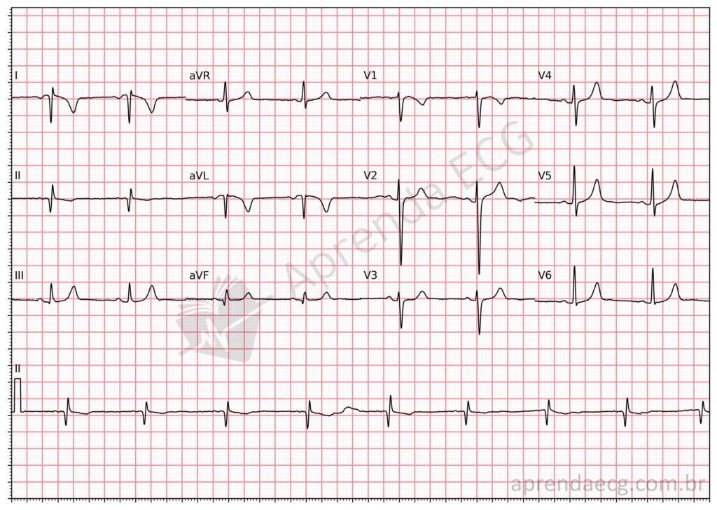
Lead reversal. Right arm–left arm reversal producing negative waves in lead I and positive waves in aVR.
Left Arm–Left Leg Reversal
- P wave in lead I is taller than in lead II (Abdollah sign)
- Inverted polarity of lead III
Right Arm–Left Leg Reversal
- Negative P wave in I, II and III
- Positive P wave in aVR
- Negative QRS complexes in the inferior wall
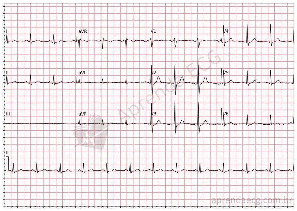
Lead reversal. Right arm–left leg reversal producing negative P waves and QRS complexes in I, II and III. The P wave is positive in aVR.
Right Arm–Right Leg Reversal
- Isoelectric line in lead II
Left Arm–Right Leg Reversal
- Isoelectric line in lead III
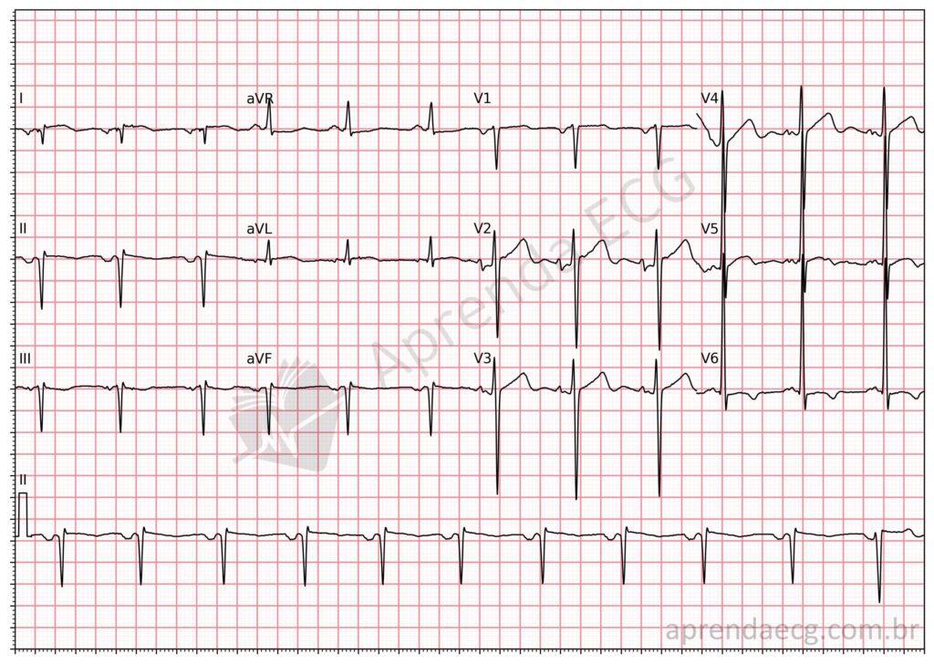
Lead reversal. Left arm–right leg reversal producing an isoelectric line in lead III.
Reversal of Upper and Lower Limb Electrodes
- Isoelectric line in lead I
Related Topics
References
- Scheffer MK, Ohe LN, de Alencar Neto JN. Manual Prático de eletrocardiograma. 2022.


