

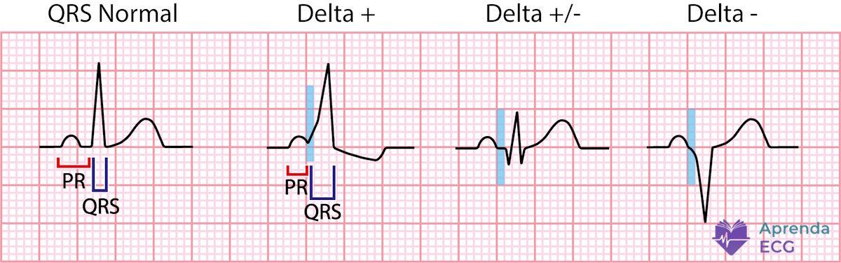
The delta wave on the ECG is the electrocardiographic manifestation of ventricular pre-excitation. It indicates that part of the ventricular myocardium is being activated earlier than it would be through physiological conduction via the atrioventricular (AV) node.
Conceptually, the delta wave is not just a morphological finding. It is the electrical expression of a specific anatomical substrate: the presence of an AV accessory pathway.
Electrophysiological Concept
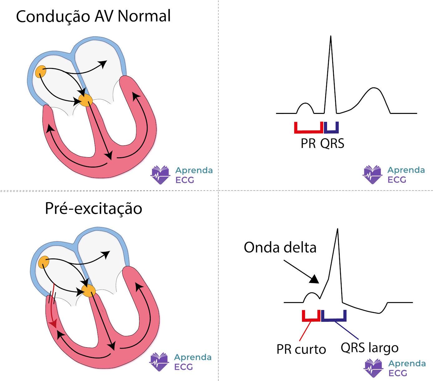
In normal conduction:
- The impulse originates in the sinus node
- It travels through the atria
- It undergoes a physiological delay in the AV node
- It descends rapidly through the His–Purkinje system
- It activates both ventricles simultaneously
In ventricular pre-excitation, an accessory pathway connects atrium and ventricle, allowing part of the impulse to “bypass” the nodal delay.
Result:
- One ventricular region is activated early
- The rest of the ventricle is activated through the normal pathway
- A fusion complex is formed
The delta wave represents exactly this slow initial activation, cell to cell, outside the His–Purkinje system.
Electrocardiographic Criteria for Ventricular Pre-excitation
Pre-excitation manifests as a set of findings:
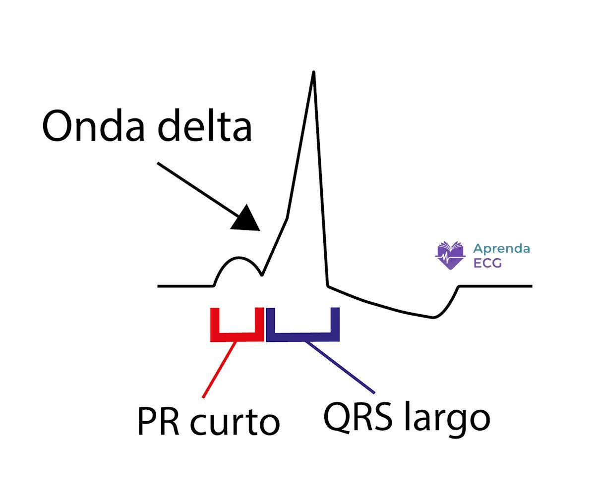
- PR interval < 120 ms
- Slurring of the initial portion of the QRS (delta wave)
- QRS ≥ 120 ms (usually)
- Secondary repolarization abnormalities
It is essential to look at the onset of the QRS in multiple leads. Analyzing a single lead in isolation is insufficient.
Why Does the PR Shorten?
The PR shortens because part of the ventricle begins to be activated before conduction through the AV node is complete.
Important:
Not every short PR means pre-excitation.
In an isolated short PR, without a delta wave, there is no evidence of early ventricular activation.
The presence of the delta wave is what confirms pre-excitation.
Degree of Pre-excitation
The expression of the delta wave depends on three main factors:
- Conduction velocity through the AV node
- Conduction velocity through the accessory pathway
- Anatomical distance from the atrial focus to each pathway
The faster the accessory pathway and the slower the nodal conduction, the greater the degree of pre-excitation.
That is why the delta wave may be:
- Obvious
- Subtle
- Intermittent
- Absent at certain times
So-called intermittent pre-excitation occurs when conduction alternates between the normal pathway and the accessory pathway.
Types of Pre-excitation
Manifest Pre-excitation
A clear delta wave on the baseline ECG.
Intermittent Pre-excitation
The delta wave disappears at certain times, usually due to changes in autonomic tone.
Concealed Accessory Pathway
Exclusively retrograde conduction.
The ECG in sinus rhythm is normal.
These pathways can take part in reentrant circuits, but they do not produce a delta wave.
Wolff-Parkinson-White Syndrome
It is essential to distinguish:
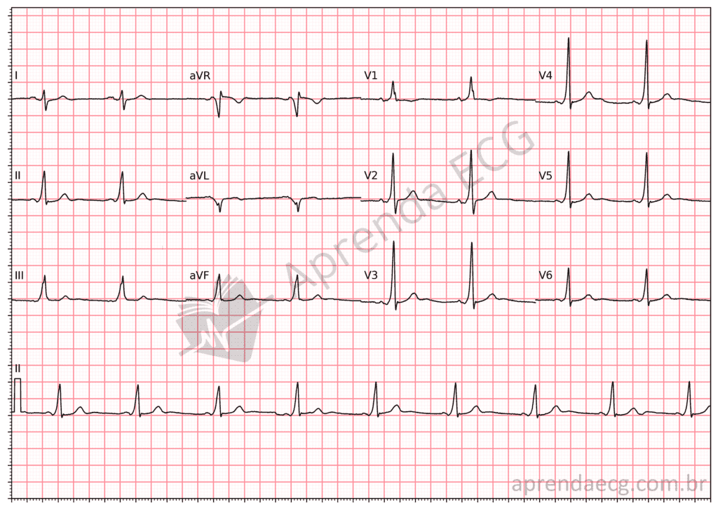
- Pre-excitation pattern: delta wave on the ECG without documented arrhythmia
- Wolff-Parkinson-White syndrome: pre-excitation + symptomatic tachyarrhythmia
Atrioventricular reentrant tachycardia (AVRT) is the most common manifestation.
During orthodromic AVRT:
- The QRS is narrow
- The delta wave disappears
In antidromic AVRT:
- The QRS is wide
- It can mimic ventricular tachycardia (VT)
Accessory Pathway Localization: the EPM Algorithm
The polarity of the delta wave allows the location of the accessory pathway to be estimated while still in sinus rhythm.
The EPM Algorithm (from the Portuguese Esquerda–Posterior–Migração: left, posterior, migration) is a practical strategy based on three sequential questions:
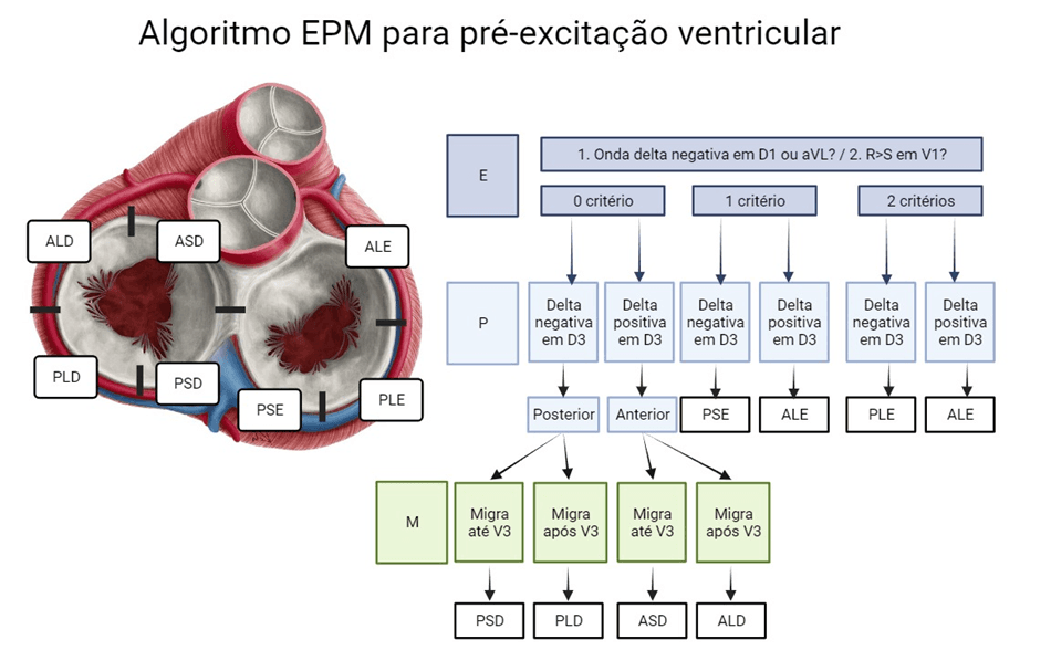
1. E (Esquerda): left or right?
Assess leads I/aVL and V1:
- Negative delta in lead I or aVL and/or R > S in V1 → suggests a left-sided pathway
- Absence of these findings → suggests a right-sided pathway
2. P: posterior or anterior?
Assess lead III:
- Negative delta → posterior
- Positive delta → anterior
3. M: precordial migration (right-sided pathways)
Assess the R-wave transition:
- Before V3 → right septal
- At V4 or later → right lateral
With these three steps, the region of the pathway can be estimated with good clinical accuracy.
The most frequent locations remain:
- Left free wall
- Posteroseptal region
For a detailed explanation, see the dedicated article on the EPM algorithm.
Delta Wave: Common Errors
- Mistaking it for bundle branch block
- Interpreting it as an old infarction
- Overvaluing an isolated short PR
- Ignoring secondary repolarization abnormalities
Systematic analysis avoids these mistakes.
Conclusion
The delta wave on the ECG is the expression of early ventricular activation via an accessory pathway.
It results from the fusion of:
- Slow initial depolarization (accessory pathway)
- Rapid subsequent depolarization (His–Purkinje)
Understanding its mechanism is more important than simply recognizing it visually.
References
- Scheffer MK, De Marchi MFN, de Alencar Neto JN, Felicioni SP. Eletrocardiograma de A a Z . São Paulo: Manole; 2024.
- Scheffer MK, Ohe LN, de Alencar Neto JN. Manual Prático de Eletrocardiograma . 2022.
- Alencar Neto JN de. Tratado de ECG . Salvador: Sanar; 2022.
- Alencar Neto JN, Sakai MH, de Almeida Neto RG, Scheffer MK, Alencar e Silva GPS, Cirenza C, et al. EPM algorithm: A stepwise approach to accessory pathway localization in ventricular pre-excitation . J Electrocardiol. 2024;84:1–8.
- Arruda MS, McClelland JH, Wang X, et al. Development and Validation of an ECG Algorithm for Identifying Accessory Pathway Ablation Site in Wolff-Parkinson-White Syndrome . J Cardiovasc Electrophysiol. 1998;9(1):2–12.


