

Systematic interpretation of the electrocardiogram (ECG) in pediatrics is challenging because of the marked physiological changes that occur during growth and because the classic diagnostic criteria were originally developed for adults. In this context, the CHILDHEART Project emerged as a pioneering initiative of the Instituto Dante Pazzanese de Cardiologia, dedicated to validating ECG criteria specific to the pediatric population. Through a series of case-control studies, the project has been establishing diagnostic scores for diseases of the right and left atria and ventricles, based on robust multivariable analyses adapted to different age groups.
In this post, we show how to apply, step by step, a practical systematic approach to pediatric ECG analysis, grounded in the evidence published by the CHILDHEART group, which aims to improve the diagnostic accuracy and clinical applicability of the ECG in children with congenital and acquired heart disease.
Key points before starting the systematic approach
- The electrocardiogram is not a gold standard; it is a surrogate. We use it as a screening tool in the initial assessment, not as a diagnosis;
- Age is indispensable for interpreting the pediatric ECG;
- The ECG will usually indicate the class of disease, not the exact malformation;
- Finally, it must be evidence-based, always using age-based normative data and the clinical and electrocardiographic context to ensure diagnostic accuracy.
First Steps of the Systematic Approach
- Step 1: What is the age?
- Step 2: Is the P wave sinus?
- Step 3: PQ interval;
- Step 4: QRS complex;
- Step 5: T wave.
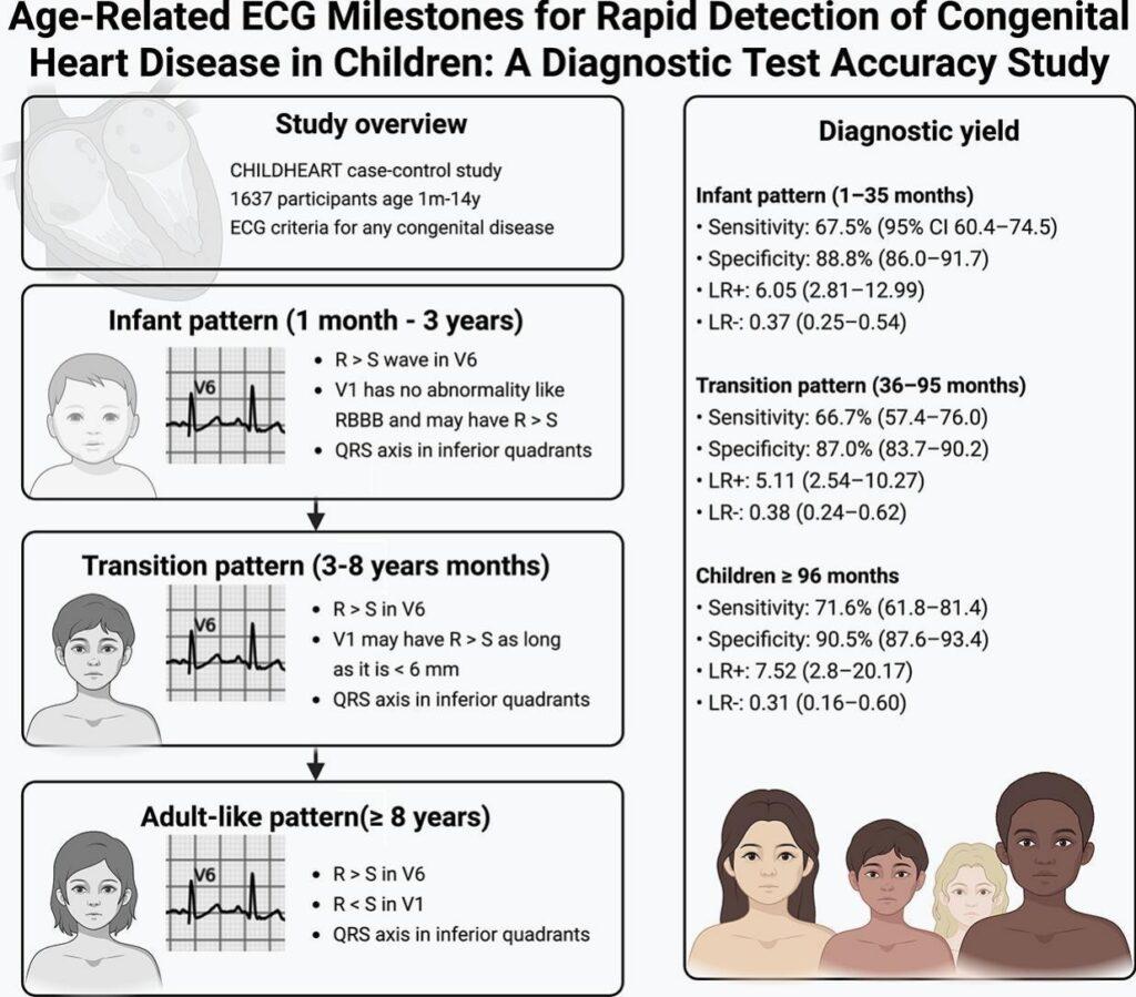
Step 1: What is the age?
Early neonatal pattern: 0 to 5 days of life
- The axis may be in the extreme right;
- R may be > S in V1;
- S may be > R in V6;
- Upright T wave in V1.
Late neonatal pattern: 5 days to 1 month
- The axis may be in the extreme right;
- R may be > S in V1;
- S may be > R in V6;
- Negative T wave in V1.
Infant pattern: 1 month to 3 years
- The axis is in the inferior quadrants (positive QRS in aVF);
- R may be > S in V1;
- R must be > S in V6.
Transitional pattern: 3 to 8 years
- The axis is in the inferior quadrants (positive QRS in aVF);
- R may be > S in V1, as long as it is not > 6 mm;
- R must be > S in V6.
Adult pattern: 8 years to adolescence
- The axis is in the inferior quadrants (positive QRS in aVF);
- R must be < S in V1;
- R must be > S in V6.
2. Is the P wave sinus?
- Upright in I and II, and plus-minus in V1.
Is there atrial disease?
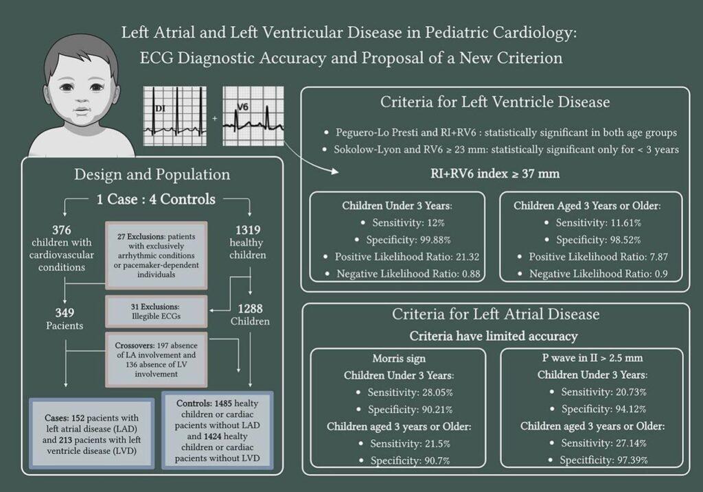
- Left atrium: use Morris (negative portion of the P wave ≥ 1 mm in amplitude and duration).
3. PQ interval
- Between 80 and 160 ms (children have a shorter PQ).
4. QRS: is there ventricular disease?
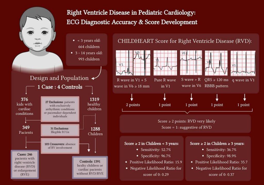
4.1. Right Ventricular Hypertrophy
| Criterion | Points |
|---|---|
| R V1 + S V6 ≥ 18 | 2 points |
| Pure R in V1 | 1 point |
| S > R in V6 | 1 point |
| QRS > 120 ms + RBBB | 1 point |
| q in V1 | 1 point |
4.2. Left Ventricular Hypertrophy
- CHILDHEART index:
- RV1 + RV6 ≥ 37 mm at any age and in either sex.
- Modified Peguero-Lo Presti:
- SV4 + SV3 ≥ 35 mm for < 3 years
- SV4 + SV3 ≥ 49 mm for ≥ 3 years
5. T wave
- The T wave may be negative in aVR, III, V1, and up to V3 (perhaps even V4).
- There must be no ST elevation or depression.
Key points
So far, CHILDHEART has not analyzed repolarization abnormalities; these are being studied for phase 2.
References
- de Alencar JN, Costa IR, Monteiro JAM, Furtado JT, Sacre RC, Guerra CB, et al. Age-related ECG milestones for rapid detection of congenital heart disease in children: A diagnostic test accuracy study. Journal of Electrocardiology. 2025.
- de Alencar JN, Monteiro JAM, Furtado JT, Sacre RC, Guerra CB, Knoblauch WS, et al. ECG Criteria and Diagnostic Score for Right Atrial Disease in Children: The CHILDHEART Study. Pediatric Cardiology. 2025.
- de Alencar JN, Monteiro JAM, Furtado JT, Sacre RC, Guerra CB, Knoblauch WS, et al. ECG criteria and diagnostic score for right ventricular disease in children: The CHILDHEART study. Journal of Electrocardiology. 2025 Sep 18;92(500):154088.
- de Alencar JN, Monteiro JAM, Furtado JT, Sacre RC, Guerra CB, Knoblauch WS, et al. ECG criteria for diagnosing left chambers disease in Pediatrics: The CHILDHEART study. Journal of Electrocardiology. 2025 Sep;92(500):154087.
- Scheffer MK, De Marchi MFN, de Alencar Neto JN, Felicioni SP. Eletrocardiograma de A a Z. São Paulo: Manole, 2024.
- Scheffer MK, Ohe LN, de Alencar Neto JN. Manual Prático de eletrocardiograma. 2022.
- Alencar JN. Tratado de ECG. 2022.


