

As the electrocardiographic analysis of acute myocardial infarction evolves, the OMI/NOMI paradigm uses the ECG as a non-dichotomous tool in its diagnostic approach, not limited to the presence or absence of ST-segment elevation in two contiguous leads.
The electrocardiographic tracing therefore expresses the dynamic changes of ischemia in different ways, and several patterns related to acute coronary occlusion (OMI) are gradually being recognized and described.
In this context, the Swirl pattern was born, described by Pendell Meyers and Stephen Smith on their famous blog (https://drsmithsecgblog.com/), first mentioned in 2013:
“The combination of precordial ST elevation and ST depression, simultaneously, should alert you to LAD occlusion. This is frequently seen when there is LV septal involvement (…), We now call this Swirl”.
In 2022, it was renamed “Precordial Swirl”. Known in Portuguese as “Divergência Precordial” (precordial swirl), it is one more OMI pattern among the more than 10 described in the literature.
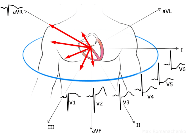
Electrocardiographic Criteria:
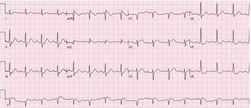
- ST elevation and/or hyperacute T wave in V1 and/or V2
- ST depression and/or T-wave inversion in V5 and/or V6
- Normal QRS (narrow, without criteria for left ventricular hypertrophy and/or intraventricular conduction blocks)
ECG in Precordial Swirl
What Does the Evidence Show?
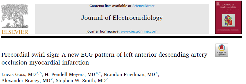
Recently published in the Journal of Electrocardiology, the article Precordial swirl sign: A new ECG pattern of left anterior descending artery occlusion myocardial infarction is the first cohort that aimed to describe, analyze, and validate this pattern. In it, the pattern was derived from the analysis of individual cases, compared with mimics (e.g., bundle branch blocks, LVH, and paced rhythm), and applied to a database of high-risk acute coronary syndrome patients, showing us that:
- The pattern is mainly associated with Left Anterior Descending artery occlusions.
- When ECGs meeting LVH criteria (in this article, specifically Sokolow-Lyon) were also included, even with a narrow QRS, the pattern's diagnostic power became null, with a positive likelihood ratio (LR+) = 1.
- When only ECGs with a normal QRS were included, the pattern still had very limited diagnostic power (LR+ = 1.38).
- The pattern more than triples its diagnostic power when the ratio between the T-wave amplitude and the S-wave amplitude of the preceding QRS in V2 (T/S ratio) is taken into account: its presence with a T/S ratio ≥ 0.4 in V2 yields an LR+ = 4.35 for OMI.
This shows the importance of proportionality between the QRS and repolarization (the ST-T complex); the article strongly emphasizes ST depression/elevation and/or a T wave that is abnormal relative to the preceding QRS.
Conclusion
Precordial Swirl emerges as an interesting OMI pattern with a predilection for the Left Anterior Descending artery. However, in this seminal article, the pattern shows a low positive likelihood ratio (LR+) and the need to differentiate it from its mimics. Its application therefore requires caution and should ideally be reserved for clinical presentations with a high pretest probability.
References
- https://drsmithsecgblog.com/septal-stemi-with-st-elevation-in-v1/
- https://drsmithsecgblog.com/the-pathophysiology-based-term-omi/
- https://drsmithsecgblog.com/precordial-swirl-20-cases-of-swirl-or/
- Goss L, Meyers HP, Friedman B, Bracey A, Smith SW. Precordial swirl sign: A new ECG pattern of left anterior descending artery occlusion myocardial infarction. J Electrocardiol. 2025;91:153931.
- Ricci F, Martini C, Scordo DM, Rossi D, Gallina S, Fedorowski A, et al. ECG Patterns of Occlusion Myocardial Infarction: A Narrative Review. Ann Emerg Med. abril de 2025;85(4):330–40.
- Scheffer MK, de Alencar Neto JN, De Marchi MFN, Felicioni SP. Eletrocardiograma na síndrome coronária aguda. São Paulo: Manole, 2025.
- Este artigo também contou com a valiosa participação de Daniel Teixeira de Campos e Barreiro


