

The N wave is a curious electrocardiographic phenomenon that may appear in patients with acute occlusion of the circumflex artery. It refers to the appearance of a notch or deflection in the terminal portion of the QRS complexes in leads II, III, and aVF or I and aVL, which changes continuously over hours to days.
Electrocardiographic Criteria
- A notch or deflection at the end of the QRS complex in leads II, III, aVF and/or the lateral leads I, aVL
- The height of the notch or deflection must be at least 2 mm relative to the PR segment.
- QRS duration in the affected leads is prolonged, with values above 120 ms.
- The N wave changes continuously over 24 hours and may disappear or turn into an S wave.
ECG Showing an N Wave
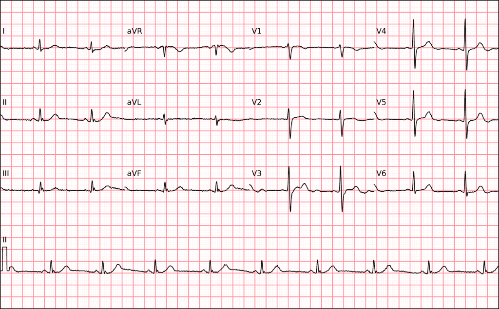
N wave. A 53-year-old woman with a hypertensive crisis and chest pain. The ECG shows a notch at the end of the QRS complex in the inferior leads. Variation in notch amplitude or its eventual disappearance within hours may indicate occlusion of the circumflex artery. The presence of an N wave in the inferior leads has a sensitivity of 77% and a specificity of 89% for LCx occlusion.
Going deeper…
The changes seen in the terminal portion of the QRS during ischemia may be caused by irregular depolarization of the inferior and left lateral regions supplied by the circumflex artery.
Because ischemia evolves, the morphology of the terminal QRS wave changes continuously over hours to days, requiring continuous monitoring, which weakens this sign for emergency diagnosis. Therefore, it does not replace other, more robust electrocardiographic and clinical criteria.
Although it is not sufficient to diagnose acute coronary occlusion (OMI), it can serve as a warning sign, especially in patients complaining of chest pain.
Related Topics
References
- Niu T, Fu P, Jia C, Dong Y, et al. The delayed activation wave in non-ST-elevation myocardial infarction. Int J Cardiol. 2013;162(2):107–11.
- Yang T, Chen J, Liu X, Xu C, et al. Non‐ST‐elevated myocardial infarction with “N” wave on electrocardiogram and culprit vessel in left circumflex has a risk equivalent to ST‐elevated myocardial infarction. Clin Cardiol. 2020;43(5):491–9.


