

An electrically inactive area is one in which ventricular activation does not occur as expected, characterized by the presence of pathological Q waves or equivalents in two contiguous leads. Q waves are usually generated as a consequence of changes produced in infarcted areas (scar/fibrosis).
Electrocardiographic Criteria
- Any Q wave in V2-V3 > 20 ms or QS complex in V2-V3
- Q wave ≥ 30 ms and ≥ 1 mm deep or QS complex in I, II, aVL, aVF or V4-V6 in two contiguous leads (I, aVL; V1-V6; II, III, aVF). The same applies to leads V7-V9
- R wave > 40 ms in V1-V2 and R/S > 1 with a positive T wave in the absence of conduction disturbances
* The criteria apply in the absence of LVH or LBBB.
Topography
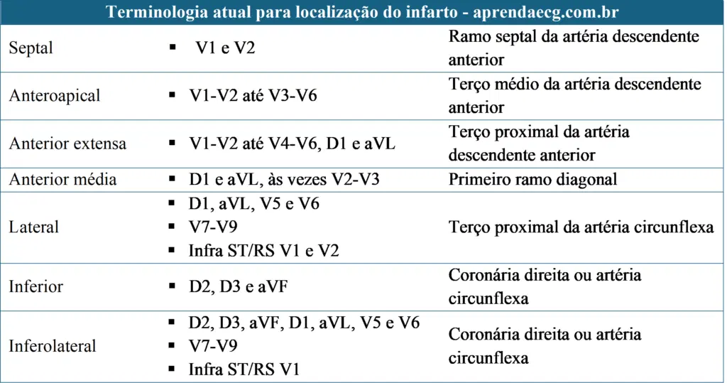
ECG in Electrically Inactive Areas
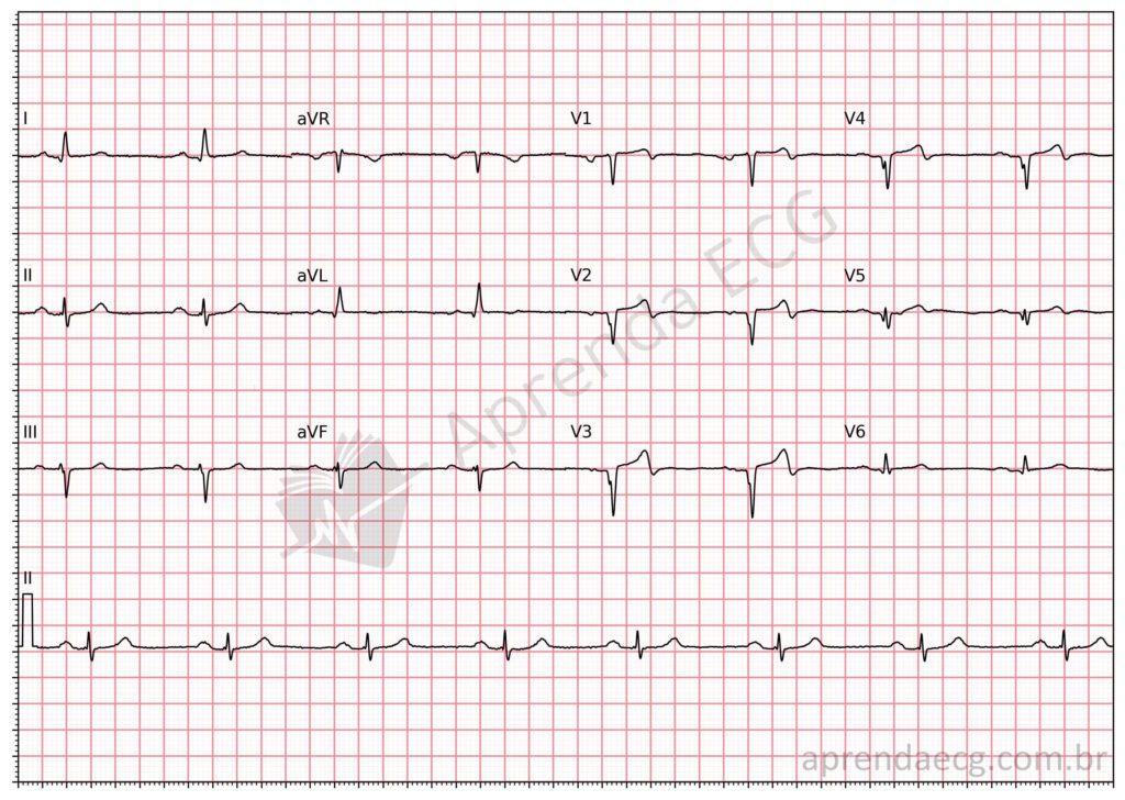
Anteroapical electrically inactive area. Q waves/QS complexes in the anteroapical wall.
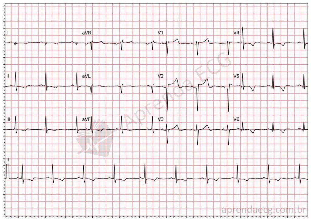
Mid-anterior electrically inactive area. Pathological Q waves in the mid-anterior wall (leads I, aVL and V2).
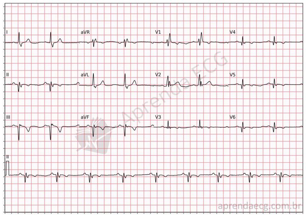
Inferolateral electrically inactive area. ECG with RBBB and pathological Q waves in the inferolateral wall (II, III, aVF, V5 and V6).
Related Topics
References
- Samesima N, God EG, Kruse JCL, Leal MG, et al. Diretriz da Sociedade Brasileira de Cardiologia sobre a Análise e Emissão de Laudos Eletrocardiográficos – 2022. Arq Bras Cardiol. 2022;93(3 Suppl 2):2–19.
- Thygesen K, Alpert JS, Jaffe AS, Chaitman BR, et al. Fourth universal definition of myocardial infarction (2018). Eur Heart J. 2018;40(3):237–69.


