

Definition
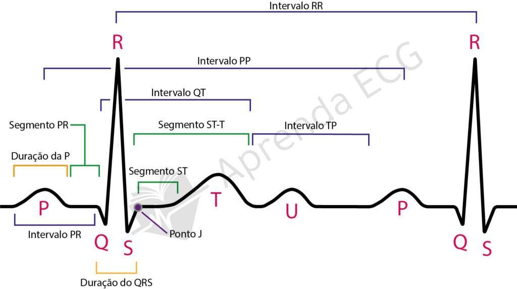
The QT interval represents the period of ventricular depolarization and repolarization, encompassing the QRS complex, the ST segment, and the T wave. Abnormalities of this interval can predispose to malignant arrhythmias, such as Torsades de Pointes.
Step by step: how to measure the QT interval
- Measure the interval from the onset of the QRS complex to the end of the T wave. Preferably use lead II or, alternatively, V5, V6, or I;
- Use a ruler or count the small squares (each square = 40 ms);
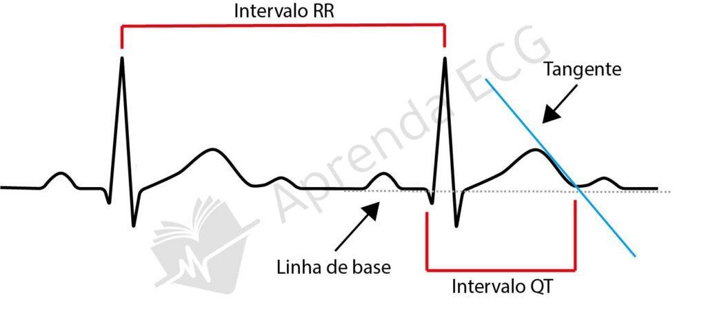
- To identify the end of the T wave, use the tangent method:
- Draw a tangent line along the downslope of the T wave;
- The point where the tangent crosses the baseline (PR segment) is considered the end of the T wave;
- Do not include the U wave in the measurement;
- Take the average of at least 3 consecutive beats in stable sinus rhythm.
QT interval behavior with heart rate
The QT interval varies inversely with heart rate, that is:
- The QT interval shortens as HR increases (tachycardia);
- The QT interval lengthens as HR decreases (bradycardia).
Corrected QT interval and its formulas
QT correction (QTc) normalizes the interval to a standard heart rate of 60 bpm. Main formulas:
- Bazett: QTcB = QT / √RR
- Most common; inaccurate outside the 60-100 bpm range;
- Widely available in medical calculators and apps.
- Fridericia: QTcFri = QT / ∛RR
- Better than Bazett for bradycardia.
- Framingham: QTcFr = QT + 0.154 x (1-RR)
- More uniform linear correction.
- Hodges: QTcH = QT + 1.75 x (HR – 60)
- Reported by some studies as more accurate, especially in the presence of a wide QRS.
- Rautaharju: QTcRa = QT x [(120 + HR) / 180]
- Showed good accuracy in recent studies, validated in large populations.
Modified QT Interval – Assessment in the Presence of a Wide QRS
In the presence of a wide QRS (bundle branch blocks, pacemaker), the QT interval is artificially prolonged because of the longer depolarization. In this setting, use the simplified Bogossian formula:
- QTm = QT – 50% of the QRS duration;
Don't forget to correct it afterward for HR using the Hodges or Fridericia formula.
Reference values
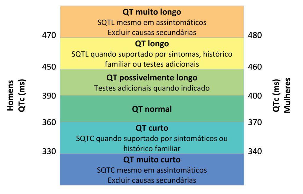
We consider the following reference values:
- Men: normal 350-450 ms
- Women: normal 360-470 ms
- QTc > 500 ms associated with a high risk of arrhythmias
- QTc < 320 ms considered short
Causes of a prolonged QT interval
- Electrolyte: hypokalemia, hypomagnesemia, hypocalcemia
- Bradyarrhythmias: sinus node dysfunction, advanced AV block
- Metabolic: hypothyroidism, hypothermia, anorexia
- Drugs: antibiotics, antiarrhythmics, antidepressants, antipsychotics, antiemetics
- Other: ischemia (Takotsubo), intracranial disease, HIV
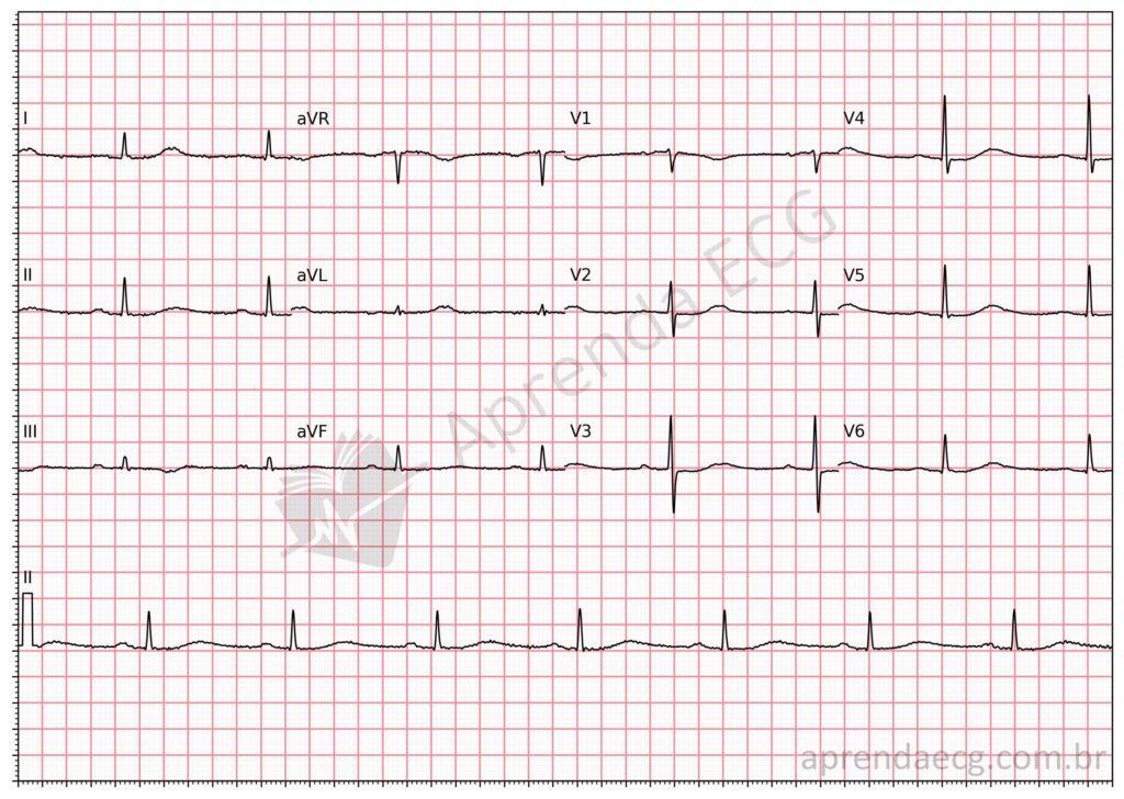
Long QT interval. The upper limit of normal for the QT interval varies with age and sex. In this example, the HR is 61 bpm, the RR interval is 980 ms, the QT interval is 498 ms, and the QTc (Bazett) is 503 ms.
Causes of a short QT interval
- Acidosis
- Digitalis effect
- Hypercalcemia
- Hyperkalemia
- Hyperthermia
- Increased vagal tone
- Myocardial ischemia
- Normal variant
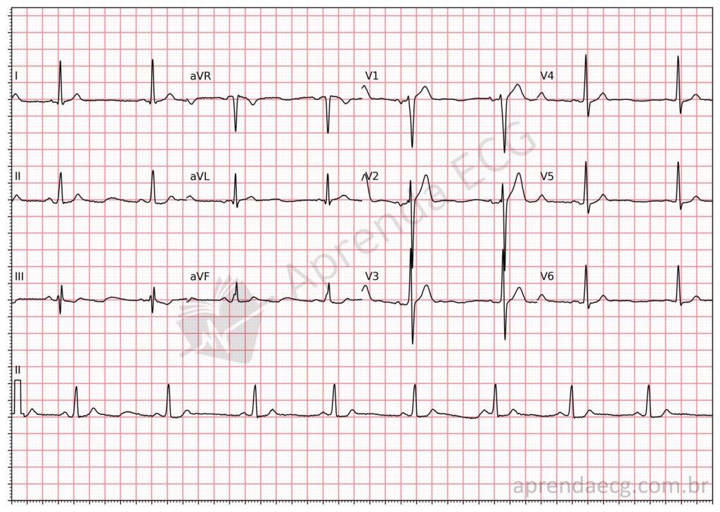
Hypercalcemia. This electrolyte disorder shortens the QT interval.
Torsades de Pointes
A potentially fatal polymorphic ventricular arrhythmia associated with a prolonged QT interval:
- Rapid (>200 bpm), paroxysmal episodes;
- Gradual inversion of the amplitude and polarity of the QRS complexes;
- Frequent association with early afterdepolarizations (tall U waves);
- May degenerate into ventricular fibrillation.
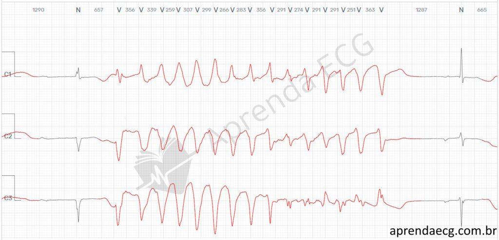
Torsade de pointes. Paroxysmal episodes of polymorphic ventricular tachycardia with gradual inversion of the QRS electrical axis, occurring in the setting of a long QT interval.
ECG Waves and Intervals
- P wave
- PR interval and segment
- QRS complex
- ST segment
- T wave
- QT interval
- U wave
- J wave
- Delta wave
- Epsilon wave
References
- Scheffer MK, De Marchi MFN, de Alencar Neto JN, Felicioni SP. Eletrocardiograma de A a Z. São Paulo: Manole, 2024.
- Scheffer MK, Ohe LN, Alencar Neto JN de. Manual Prático de Eletrocardiograma do Setor de Emergências do Instituto Dante Pazzanese de Cardiologia. São Paulo: Editora dos Editores; 2022. 426 p.
- Postema PG, De Jong JSSG, Van der Bilt IAC, Wilde AAM. Accurate electrocardiographic assessment of the QT interval: Teach the tangent. Hear Rhythm. 2008;5(7):1015–8.
- Bazett HC. An Analysis of the Time-Relations of Electrocardiograms. Ann Noninvasive Electrocardiol. 1997;2(2):177–94.
- Fridericia LS. The duration of systole in an electrocardiogram in normal humans and in patients with heart disease. Ann Noninvasive Electrocardiol. 2003;8(4):343–51.
- Sagie A, Larson MG, Goldberg RJ, Bengtson JR, et al. An improved method for adjusting the QT interval for heart rate (the Framingham Heart Study). Am J Cardiol. 1992;70(7):797–801.
- Hodges M, Salerno D, Erlien D. Bazett’s QT correction reviewed: evidence that a linear correction is better (Abstr). J Am Coll Cardiol. 1983;(1):6944.
- Rautaharju PM, Mason JW, Akiyama T. New age- and sex-specific criteria for QT prolongation based on rate correction formulas that minimize bias at the upper normal limits. Int J Cardiol. 2014;174(3):535–40.
- Bogossian H, Linz D, Heijman J, Bimpong-Buta N-Y, et al. QTc evaluation in patients with bundle branch block. IJC Hear Vasc. 2020;30:100636.
- Bogossian H, Frommeyer G, Ninios I, Hasan F, et al. New formula for evaluation of the QT interval in patients with left bundle branch block. Hear Rhythm. 2014;11(12):2273–7.
- Erkapic D, Frommeyer G, Brettner N, Sözener K, et al. QTc interval evaluation in patients with right bundle branch block or bifascicular blocks. Clin Cardiol. 2020;43(9):957–62.
- Viskin S. The QT interval: Too long, too short or just right. Hear Rhythm. 2009;6(5):711–5.
- Kahlon SS, Sikandar R, Tejovath S, Nair S, et al. Diagnosing Torsades De Pointes Based on Correlation to QT Interval: A Systematic Review. Cureus. 2022;14(8):1–9.
- Dave J. Torsade de Pointes. Medscape. 2022.


