

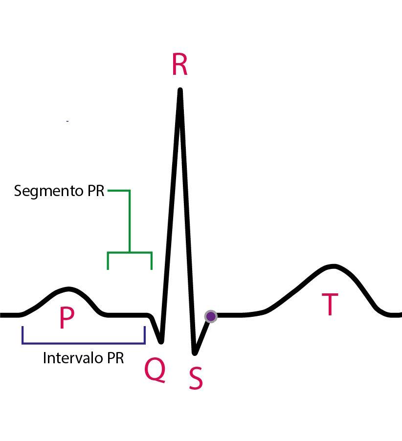
The PR interval extends from the onset of the P wave to the onset of the QRS complex and represents the electrical conduction time from the atria to the ventricles. Within it there is a specific portion called the PR segment, which corresponds only to the isoelectric stretch after the end of the P wave.
Although they share the same landmark, interval and segment are not synonyms. The former includes all atrioventricular conduction measurable on the ECG; the latter represents only the isoelectric phase related to nodal delay.
In this schematic summary, we will review the definition of each, the normal values, and the main associated abnormalities, focusing on practical application during systematic ECG reading.
PR Interval
Definition
Interval extending from the onset of the P wave to the onset of the QRS complex.
Includes:
- Atrial depolarization
- Intra-atrial conduction
- Nodal delay in the AV node
- Conduction through the proximal His bundle
What It Represents
Reflects the total atrioventricular conduction time.
The main determinant of its value is the physiological delay in the AV node, which is modulated by the autonomic nervous system. For this reason, the PR interval varies inversely with heart rate.
Normal Values
- Normal (adults): 120-200 ms (3 to 5 small boxes)
- < 120 ms: short PR
- > 200 ms: prolonged PR
Age-related variation:
- Children: the PR interval tends to be shorter, ranging approximately from 90 to 170 ms, depending on age and heart rate.
- Older adults: there may be slight physiological PR prolongation, related to greater fibrosis of the conduction system and a higher prevalence of degenerative nodal disease.
In addition to age, the PR interval is influenced by heart rate and autonomic tone.
Main Abnormalities
Prolonged PR (>200 ms)

Main mechanisms/causes:
- Increased vagal tone
- Drugs that depress nodal conduction (beta-blockers, verapamil, diltiazem, digoxin)
- Degenerative disease of the conduction system
- Ischemia or inflammation involving the AV node
Progressively lengthening PR with a blocked QRS

Fixed PR with sudden blocked QRS complexes

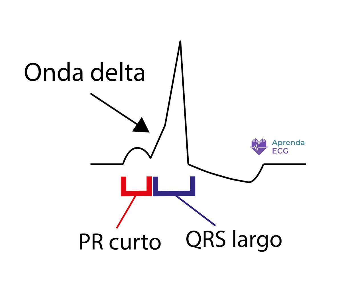
Short PR (<120 ms)
- Ventricular preexcitation (e.g., Wolff-Parkinson-White)
- Junctional rhythm
- Low atrial rhythms
- Enhanced conduction through the atrioventricular node
PR Segment
Definition
Isoelectric stretch between the end of the P wave and the onset of the QRS complex.
Corresponds to the period when there is no atrial electrical activity detectable at the body surface, predominantly reflecting nodal and early infranodal conduction.
What It Represents
Represents the AV nodal delay itself.
Serves as the baseline reference for ST-segment analysis.
During sinus tachycardia, it may show a slight downward slope, with no pathological significance on its own.
Normal Values
Should be isoelectric relative to the TP segment.
Small variations depend on how the baseline is defined and on heart rate.
Main Abnormalities
Diffuse PR-segment depression
Focal PR deviation
- May occur in atrial injury
- May accompany localized inflammatory processes
PR-segment deviation should not be interpreted in isolation; its analysis should always consider the clinical context and concomitant ST-segment changes.
References
- Scheffer MK, De Marchi MFN, de Alencar Neto JN, Felicioni SP. Eletrocardiograma de A a Z. São Paulo: Manole; 2024.
- Surawicz B, Knilans TK. Chou's electrocardiography in clinical practice. 6th ed. Philadelphia: Saunders Elsevier; 2008.


