

Atrioventricular (AV) blocks are delays in the conduction of the impulse from the atria to the ventricles. Anatomically, these delays may be located in the AV node itself (nodal block), in the His-Purkinje trunk (intra-Hisian block), or below it (infra-Hisian block). They can also be classified as first-, second-, or third-degree.
ECG Criteria
First-Degree AV Block
- PR interval > 200 ms
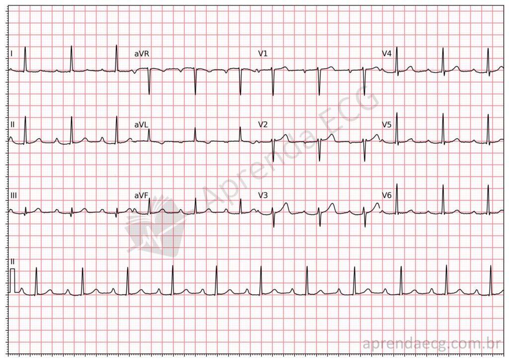
First-degree AV block. Characterized by delayed AV conduction, expressed on the electrocardiogram as a PR interval prolonged beyond 200 ms.
Second-Degree AV Block Type 1
- Progressive lengthening of the PR interval until AV conduction is blocked and a sinus beat fails to conduct
- The PR interval after the blocked P wave is shorter than the PR interval preceding the block
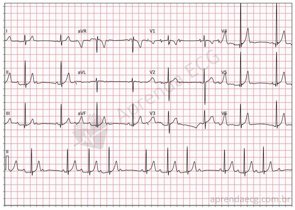
Second-degree Mobitz 1 AV block. Also known as the Wenckebach phenomenon, it refers to progressive lengthening of the PR interval until a blocked P wave occurs. The PR interval following the pause caused by the block is shorter than the PR interval preceding it.
Second-Degree AV Block Type 2
- Sudden interruption of AV conduction, seen as a blocked P wave
- The PR interval is fixed
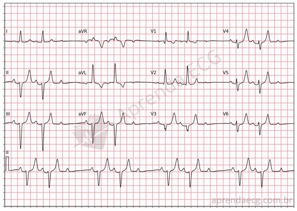
Second-degree Mobitz 2 AV block. In this form of block, the PR interval is fixed and the blocked P wave appears intermittently on the tracing.
Second-Degree 2:1 AV Block
- Alternation of a conducted sinus P wave and a blocked one
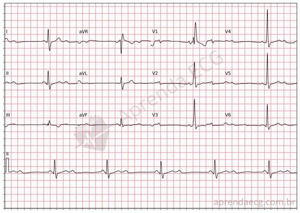
2:1 AV block. In 2:1 AV block, one P wave conducts and is followed by a QRS complex, and the next is blocked, producing a pause.
Advanced Second-Degree AV Block
- AV conduction occurs in fewer than 50% of sinus beats, at a ratio of 3:1, 4:1, or higher
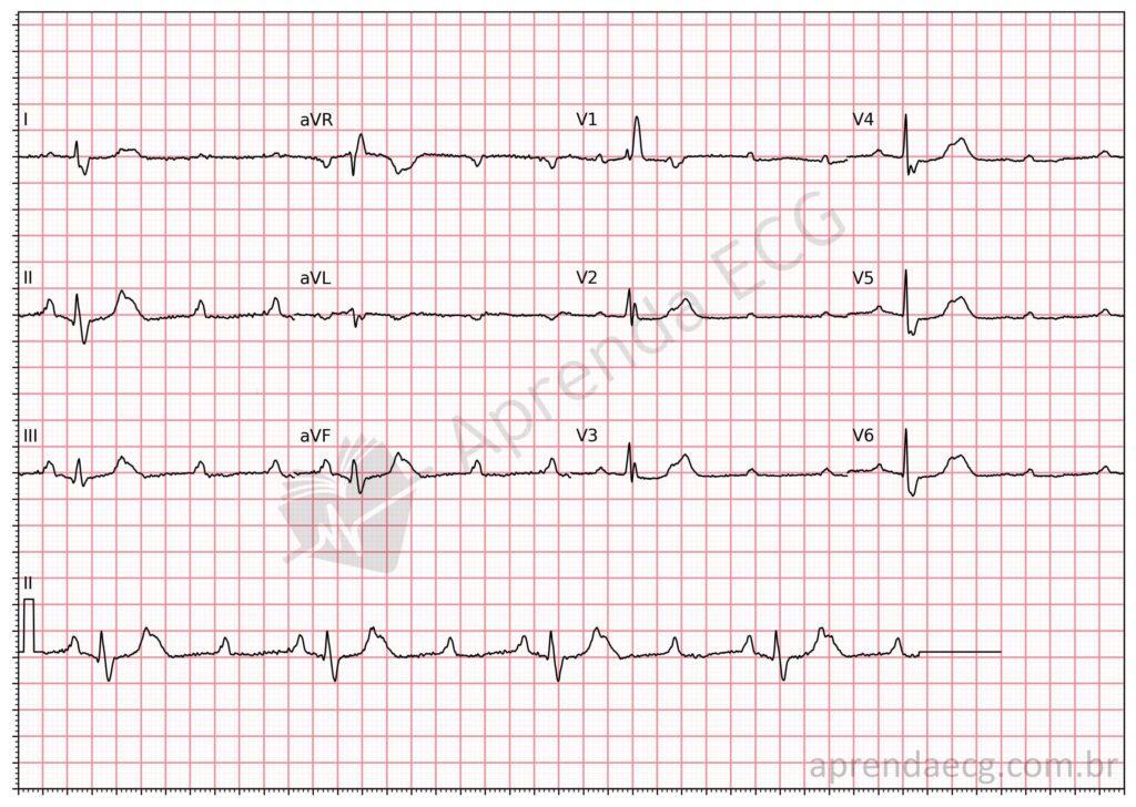
Advanced AV block. In this form of block, AV conduction is 3:1 or higher. In this example, note 2 blocked P waves and one P wave that conducts, producing a QRS complex. Note that the PR interval of the conducted P wave is fixed, which distinguishes it from complete heart block.
Third-Degree (Complete) AV Block
- There is no association between atrial and ventricular electrical activity (AV dissociation)
- The atrial rate is faster than that of the escape rhythm
- QRS complexes may be narrow (junctional escape rhythm) or wide (ventricular escape rhythm)
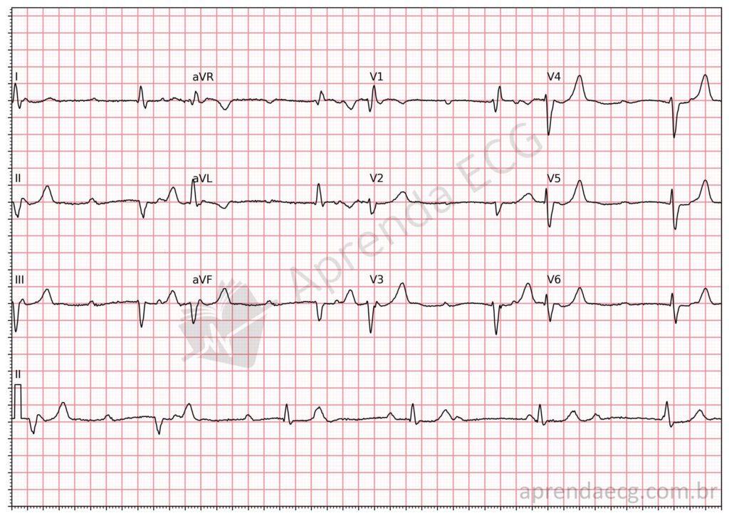
Third-degree (complete) AV block. There is complete dissociation between atrial and ventricular activity. P waves and QRS complexes are independent and bear no relationship to each other.
Related Topics
References
- Scheffer MK, De Marchi MFN, de Alencar Neto JN, Felicioni SP. Eletrocardiograma de A a Z. São Paulo: Manole, 2024.
- Samesima N, God EG, Kruse JCL, Leal MG, et al. Diretriz da Sociedade Brasileira de Cardiologia sobre a Análise e Emissão de Laudos Eletrocardiográficos – 2022. Arq Bras Cardiol. 2022;93(3 Suppl 2):2–19.
- Kusumoto FM, Schoenfeld MH, Barrett C, Edgerton JR, et al. 2018 ACC/AHA/HRS Guideline on the Evaluation and Management of Patients With Bradycardia and Cardiac Conduction Delay. J Am Coll Cardiol. 2019;74(7):e51–156.
- Issa Z, Miller J, Zipes D. Clinical Arrhythmology and Electrophysiology: A Companion to Braunwald’s Heart Disease. 3a ed. Philadelphia: Elsevier; 2019. 752 p.


