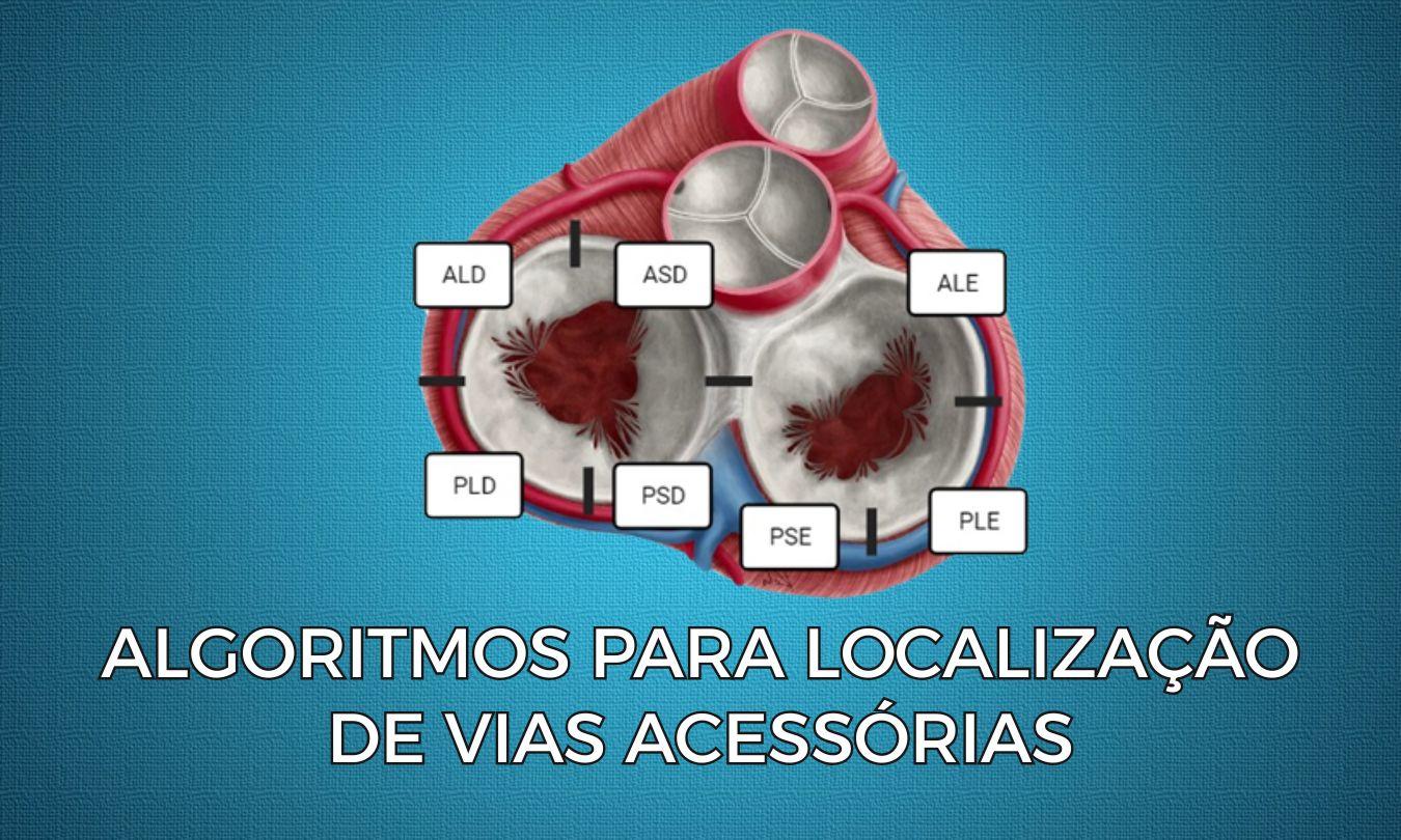
Accurately localizing accessory pathways in patients with ventricular preexcitation or Wolff-Parkinson-White (WPW) syndrome is a frequent challenge in electrophysiology. Although widely used algorithms exist, such as Arruda and EASY, the new EPM algorithm offers a systematic and didactic approach that makes it easier to identify and treat these anomalous conduction pathways. In this post, we explore how this algorithm works and its main results.
Why Correct Accessory Pathway Localization Matters
In WPW syndrome, precise identification of the accessory pathway location is crucial for the success of therapeutic procedures such as catheter ablation. Ablation guided by correct localization of the accessory pathway allows effective interruption of ventricular preexcitation, preventing associated complications such as supraventricular arrhythmias.
The EPM Algorithm: How Does It Work?
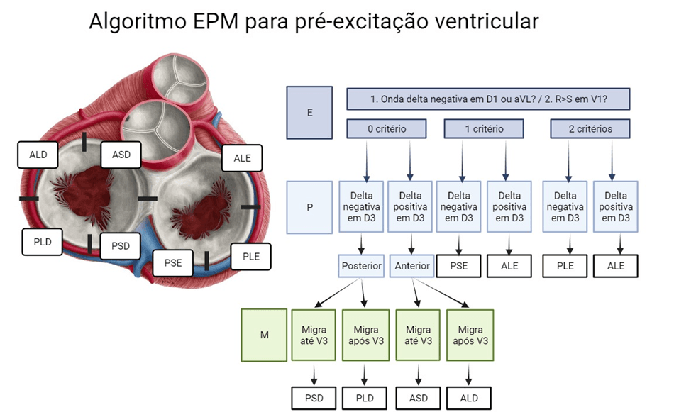
The EPM algorithm was developed to provide a logical, structured method for localizing accessory pathways on the electrocardiogram (ECG). It follows three main steps, which can be remembered by the acronym EPM (from the Portuguese initials):
- Step “E” (Left or Right?): in this step, the goal is to determine whether the accessory pathway is on the left or right side of the heart. Two features are assessed:
If both features are present, the pathway is left-sided; if only one is present, the pathway is left septal. The absence of both signs suggests a right-sided accessory pathway.- Presence of a negative delta wave in lead I or aVL. Presence of an R wave larger than the S wave in V1.
- Step “P” (Anterior or Posterior Location?): the polarity of the delta wave in lead III is analyzed. A negative delta wave indicates a posterior pathway; a positive one indicates an anterior pathway.
- Step “M” (Migration of Precordial Polarity): exclusive to right-sided pathways. A polarity change before V3 indicates a right septal pathway, whereas a change after V4 indicates a right lateral pathway.
Comparison with Traditional Algorithms
The study that evaluated EPM compared its diagnostic accuracy with the Arruda and EASY algorithms. The results showed that EPM had a diagnostic accuracy of 51.45%, while the Arruda algorithm reached 53.29% and EASY 44.69%. Although these figures may seem modest, it is important to note that EPM showed excellent interobserver agreement, with kappa statistics above 0.9, indicating high consistency between different examiners.
Sensitivity and Specificity of the EPM Algorithm
EPM showed a sensitivity of 95.73% for identifying left- versus right-sided pathways and a specificity of 74.33%. These values are comparable to those of widely used algorithms, making EPM a promising tool for clinical practice.
Application Examples
In the Tools section of our website, you will find practical examples of how to apply the EPM algorithm.
Conclusion
The EPM algorithm offers an effective, intuitive step-by-step approach to accessory pathway localization, on par with traditional algorithms and with the advantage of high consistency between different operators. Its use in clinical practice can optimize pathway localization and improve ablation outcomes. Given these promising results, EPM stands out as a robust tool in the electrophysiology arsenal.
References
- de Alencar Neto JN, Sakai MH, de Almeida Neto RG, Scheffer MK, Alencar e Silva GPS, Cirenza C, et al. EPM algorithm: A stepwise approach to accessory pathway localization in ventricular pre-excitation. J Electrocardiol. 2024;84(500):1–8.