
The EPM algorithm was developed to provide a logical, structured method for localizing accessory pathways on the electrocardiogram (ECG). It follows three main steps, which can be remembered by the acronym EPM (from the Portuguese initials). The algorithm uses the polarity of the delta wave in the frontal plane leads and the QRS transition in the precordial leads to localize the accessory pathways:
- Step 1 (“E”, from esquerda, left): determines whether the accessory pathway is left- or right-sided
- Step 2 (“P”, posterior): establishes whether the accessory pathway has a posterior location
- Step 3 (“M”, migration): migration of precordial polarity (only for right-sided accessory pathways)
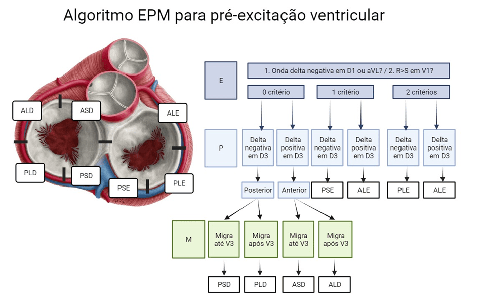
Examples of Applying the EPM
Example 1
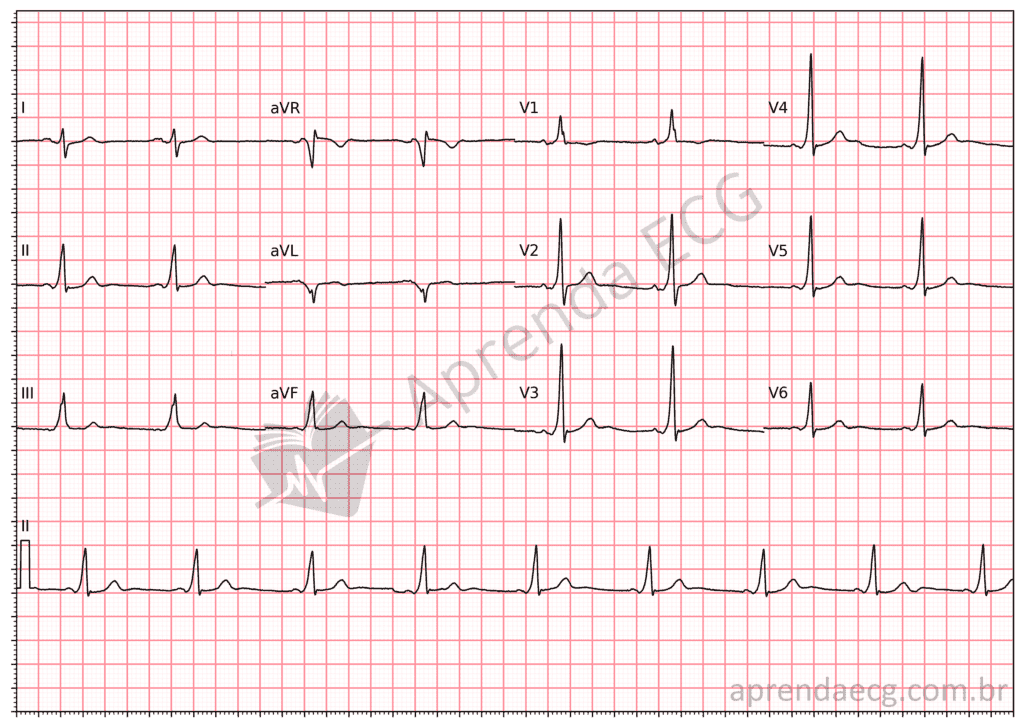
In step E we have a negative delta wave in aVL and an R wave larger than the S wave in V1. In addition, the delta wave is positive in III (step P). Therefore, this is a left anterolateral (LAL) pathway.
Example 2
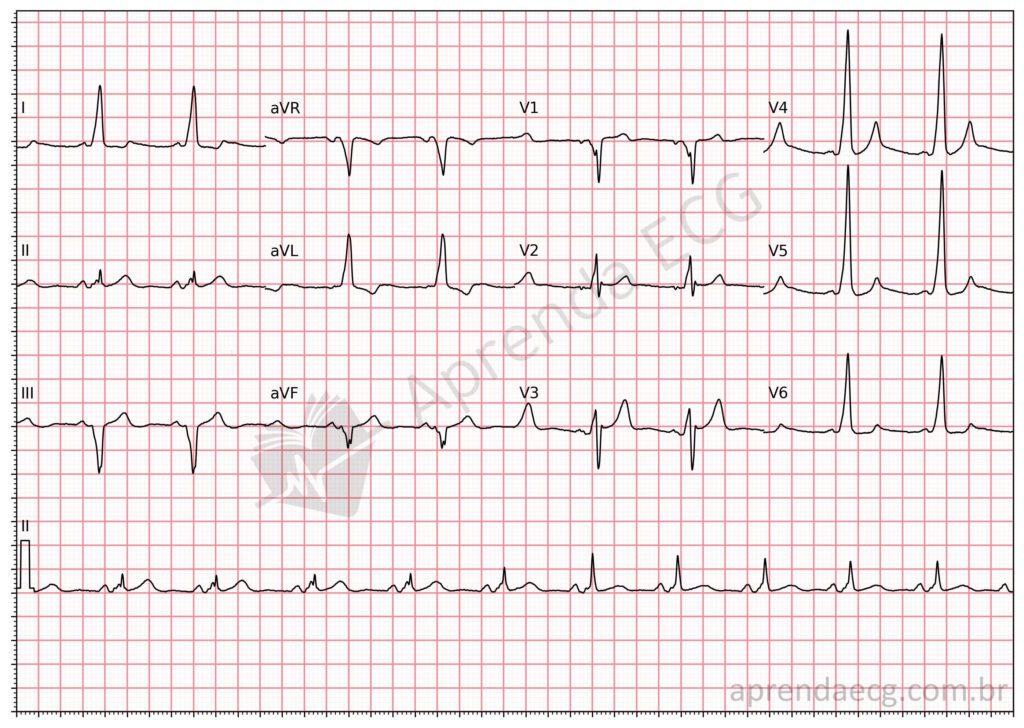
In step E, none of the criteria are met, since the delta wave is positive in I and aVL and the S wave is larger than the R wave in V1. In step P, we see a negative delta wave in III, and in step M, the precordial polarity migrates after V3; thus, we have a right posteroseptal (RPS) pathway.
References
- de Alencar Neto JN, Sakai MH, de Almeida Neto RG, Scheffer MK, Alencar e Silva GPS, Cirenza C, et al. EPM algorithm: A stepwise approach to accessory pathway localization in ventricular pre-excitation. J Electrocardiol. 2024;84(500):1–8.