Wide QRS tachycardias are among the most challenging scenarios in clinical electrocardiography. By definition, they are characterized by a heart rate ≥ 100 bpm with QRS complexes ≥ 120 ms. However, the real problem is not the width of the QRS, but the origin of the tachycardia.
For teaching purposes, three mechanisms account for the vast majority of cases:
- Ventricular tachycardia (VT): responsible for about 80% of wide QRS tachycardias, especially in the presence of structural heart disease. The impulse originates in the ventricle and spreads predominantly through slow myocardial conduction.
- Supraventricular tachycardia with aberrancy (SVT-A): accounts for approximately 15–25% of cases. The rhythm is supraventricular, but intraventricular conduction occurs with a preexisting or functional bundle branch block.
- Preexcited supraventricular tachycardia (SVT-P): less common (1–6%), it occurs when the atrial impulse is conducted anterogradely over an accessory pathway, initially activating the ventricle outside the His-Purkinje system.
The distinction between VT and SVT-A is usually the classic focus of diagnostic algorithms. However, the real electrocardiographic sticking point is differentiating VT from SVT-P, because both share a central element: initial ventricular activation through slow myocardial conduction.
The real problem is not the wide QRS
In practice, most wide QRS tachycardias are VT, especially in patients with structural heart disease. This means that the pretest probability is already high even before detailed morphological analysis.
A common conceptual error is to treat the ECG as a binary test capable of “deciding” the diagnosis on its own. However, ECG criteria modify probabilities; they do not create absolute certainty.
When reasoning ignores the clinical context and anchors exclusively on an algorithm, it creates a false sense of diagnostic security.
Why do traditional algorithms fail in SVT-P?
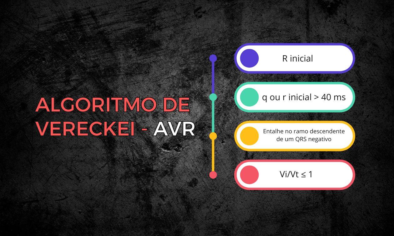
Most classic algorithms, such as the Brugada algorithm and Vereckei's aVR-based algorithm, were developed to differentiate VT from SVT with aberrancy.
The rationale for these methods is sound: in SVT-A, initial activation occurs through the His-Purkinje system; in VT, it occurs through slow myocardial conduction. The problem arises when we analyze SVT-P.
In supraventricular tachycardia conducted over an accessory pathway, initial ventricular activation also occurs outside the specialized conduction system. Electrophysiologically, the onset of the QRS in SVT-P resembles that of VT.
Practical consequence: many classic criteria classify SVT-P as VT.
The only truly specific exception remains unequivocal atrioventricular dissociation, a finding highly specific for VT but poorly sensitive.
Therefore, the limitation is not only methodological; it is structural. The ECG is trying to distinguish two phenomena that share the same initial electrophysiological principle.
Attempts to break the deadlock
Recognizing this limitation, some authors proposed algorithms specifically designed to differentiate VT from SVT-P.
Steurer Algorithm
Proposed in 1994, it was the first systematic attempt to tackle this distinction directly. Structured in three sequential steps, it prioritizes criteria with very high specificity for VT. It showed 100% specificity but limited sensitivity (~75%). In other words, it confirms VT reliably but does not adequately rule it out.
Modified Vereckei Algorithm
Published in 2023, the modification proposed by Vereckei added a criterion in lead aVR that explores the initial direction of ventricular activation. Sensitivity and overall accuracy improved compared with the Steurer algorithm, but at the cost of reduced specificity.
It is an incremental improvement, not a definitive solution.
Serial Prelocalization Algorithm
More recently, an algorithm was proposed based on the concept of prelocalizing the initial site of ventricular activation. The strategy organizes reasoning into two sequences: first it differentiates VT from SVT-A; then it distinguishes VT from SVT-P.
In the initial studies, it outperformed the classic algorithms, with high sensitivity and accuracy. However, the data come from retrospective, single-center cohorts and still lack broad external validation.
Once again, we see conceptual progress, but not the elimination of uncertainty.
Limitations of the ECG in this diagnosis
Even with refined criteria and sequential algorithms, the electrocardiogram has inherent limitations in differentiating VT from SVT-P.
First, the surface ECG is an indirect representation of the heart's three-dimensional electrical activation. Small variations in the site of origin, the presence of myocardial scar, or ventricular anatomy can produce similar morphological patterns for different mechanisms.
Second, tracing quality directly influences interpretation. Low QRS amplitude, artifacts, excessive filtering, or improper electrode placement can alter critical morphologies, especially for criteria that depend on fine analysis of the initial portion of the QRS.
Third, the absence of a baseline ECG limits essential morphological comparisons. Without knowing the prior intraventricular conduction pattern, the reader loses one of the most useful tools for putting the tachycardia in context.
In addition, some clinical situations distort the classic principles:
- Extensive cardiomyopathies with diffuse fibrosis.
- Metabolic disturbances, such as severe hyperkalemia.
- Use of sodium channel–blocking drugs.
- Idiopathic VTs arising near the His-Purkinje system.
In these scenarios, ventricular activation may mimic the patterns expected in SVT or, conversely, produce atypical morphologies that defy any algorithm.
Finally, there is interobserver variability. Interpreting notches, relative wave areas, or initial activation times depends on experience and, in many cases, on subjective judgment.
These limitations do not invalidate the ECG, but they call for diagnostic humility. The test is powerful, but not omniscient.
What this teaches about ECG reasoning
The persistence of multiple algorithms over decades reveals an important point: if a definitive method existed, the others would have disappeared.
The differential diagnosis of wide QRS tachycardias is not a purely morphological problem. It is probabilistic.
Some principles remain solid:
- Most wide QRS tachycardias are VT.
- Structural heart disease dramatically increases the probability of VT.
- Unequivocal AV dissociation virtually confirms VT.
- No single algorithm achieves perfect accuracy.
In the acute setting, especially in the face of hemodynamic instability, guidelines recommend treating every wide QRS tachycardia as VT until proven otherwise.
After stabilization, algorithms become useful tools for diagnostic refinement and treatment planning, not substitutes for clinical judgment.
Acknowledging uncertainty is not a diagnostic weakness. It is electrophysiological maturity.
Conclusion
Differentiating VT from SVT-P remains one of the most complex challenges in electrocardiography.
Traditional algorithms were designed to distinguish VT from SVT with aberrancy and have a structural limitation when faced with SVT-P. More recent attempts have brought incremental improvements, but no definitive solution.
Given this, the safest approach remains integrating clinical probability, critical ECG analysis, and therapeutic prudence.
In wide QRS tachycardia, the ECG helps a lot, but it rarely decides on its own.
References
- Issa ZF, Miller JM, Zipes DP. Clinical Arrhythmology and Electrophysiology: A Companion to Braunwald's Heart Disease. 3rd ed. Philadelphia: Elsevier; 2019.
- Akhtar M, Shenasa M, Jazayeri M, Caceres J, Tchou PJ. Wide QRS complex tachycardia. Reappraisal of a common clinical problem. Ann Intern Med. 1988;109(11):905–12. DOI: 10.7326/0003-4819-109-11-905
- Kashou AH, Noseworthy PA, DeSimone CV, Deshmukh AJ, Asirvatham SJ, May AM. Wide Complex Tachycardia Differentiation: A Reappraisal of the State-of-the-Art. J Am Heart Assoc. 2020;9(11):e016598. DOI: 10.1161/JAHA.120.016598
- Vereckei A. Current algorithms for the diagnosis of wide QRS complex tachycardias. Curr Cardiol Rev. 2014;10(3):262–76. DOI: 10.2174/1573403X10666140514103309
- Steurer G, Gürsoy S, Frey B, Simonis F, Andries E, Brugada P, et al. The differential diagnosis on the electrocardiogram between ventricular tachycardia and preexcited tachycardia. Clin Cardiol. 1994;17(6):306–8. DOI: 10.1002/clc.4960170606
- Vereckei A, Simon A, Szénási G, Miller JM. The Application of a New, Modified Algorithm for the Differentiation of Regular Ventricular and Pre-Excited Tachycardias. Heart Lung Circ. 2023;32(6):719–25. DOI: 10.1016/j.hlc.2023.03.016
- Ni H, Wang Q, Wang Z, Xu W, Pan X, Zhu J, et al. Study on the feasibility of distinguishing ventricular and pre-excited arrhythmia rhythms by a new algorithm. J Electrocardiol. 2024;87:153817. DOI: 10.1016/j.jelectrocard.2024.153817
- Ni H, Huang Y, Pan X, Zhang X, Wan Z, Zhai C, et al. Effective discrimination of wide QRS complex tachycardia with a new algorithm - the Prelocalization Series Algorithm. BMC Cardiovasc Disord. 2025;25:134. DOI: 10.1186/s12872-025-04583-1