AV nodal reentrant tachycardia (AVNRT) and orthodromic atrioventricular reentrant tachycardia (AVRT) are easily confused, and not only because of their names. Their electrocardiographic manifestations also share similarities that can make the diagnosis challenging. That is why it is essential to revisit the main concepts of these supraventricular tachycardias and highlight their differences, ensuring accurate ECG interpretation and more effective clinical management.
Review
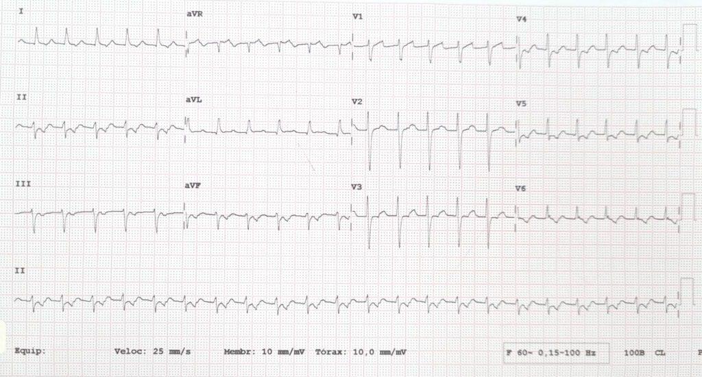
AVNRT occurs when there are two conduction pathways through the AV node, a slow pathway responsible for anterograde activation and a fast pathway responsible for retrograde activation. Its main electrocardiographic features are:
- The retrograde P wave may be hidden within the QRS complex or appear right after the QRS, forming a pseudo-S wave in II, III and aVF and a pseudo-r' in V1
- Short RP interval, < 70 ms
- Regular RR interval
- HR usually between 150 and 250 bpm
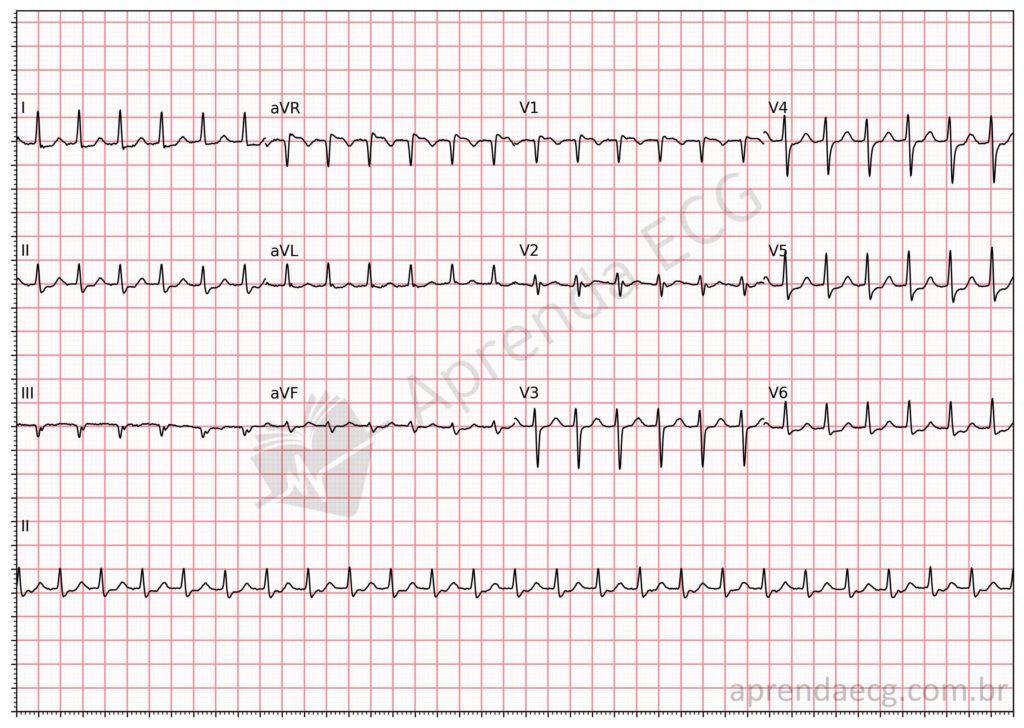
Orthodromic AVRT, on the other hand, results from reentry involving the conduction system and an accessory pathway. In orthodromic AVRT, the accessory pathway forms the retrograde limb of the circuit, while the conduction system forms the anterograde limb. Its main electrocardiographic features are:
- Narrow QRS tachycardia with a regular RR interval
- Short RP interval, > 70 ms
- HR between 150 and 250 bpm
- ST-segment depression may occur because the ST segment fuses with the retrograde P wave
- Electrical alternans (QRS alternans)
Electrocardiographic Differences
In clinical practice, even when the electrocardiographic criteria are known, distinguishing these two supraventricular arrhythmias can be challenging. However, analysis of the RP interval (the distance between the R wave and the next retrograde P wave) can provide valuable clues. In orthodromic AVRT, this interval is usually longer than 70 ms and is often accompanied by ST-segment depression, an additional criterion that helps with the distinction. In AVNRT, by contrast, the RP interval is typically shorter than 70 ms, which helps point toward the diagnosis.
References
- Scheffer MK, De Marchi MFN, de Alencar Neto JN, Felicioni SP. Eletrocardiograma de A a Z. São Paulo: Manole, 2024.
- Alencar JN. Tratado de ECG. 2022.