Have you ever analyzed an ECG and found an interatrial block (IAB)? We know this finding is crucial and well documented in the literature (including here on Aprenda ECG!). But what does it really mean for the patient?
It is not just an isolated report. It is time to connect this electrocardiographic finding to its clinical consequences. This is exactly where the discussion of Bayés syndrome comes in: it is the key to understanding the real impact of IAB and, more importantly, to predicting possible complications. Keep reading and find out how a simple IAB can hide a much greater risk.
Review of Interatrial Blocks
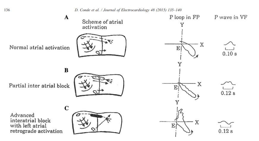
- First-degree (partial) IAB: characterized by a P wave lasting 120 ms or more, usually with biphasic morphology in leads such as II, III and aVF. Conduction is delayed but not completely blocked.
- Second-degree IAB: in this case, interatrial conduction may be normal or intermittently show features of first- or third-degree IAB on the same ECG or at different times. It is known as “atrial aberrancy.”
- Third-degree (advanced) IAB: the impulse is blocked in the region of Bachmann's bundle and the fossa ovalis, allowing conduction only through the coronary sinus. The P wave is prolonged and has a plus-minus morphology in the inferior leads.
Bayés Syndrome
Conde and Baranchuk proposed the term “Bayés syndrome” and revolutionized the way clinicians interpret the electrocardiographic finding of “interatrial block.” The basis of Bayés syndrome is fibrotic atrial cardiomyopathy, which leads to left atrial hypocontractility. This dysfunction causes blood stasis and promotes atrial remodeling, which can lead to supraventricular arrhythmias such as atrial fibrillation (AF). As a result, a hypercoagulable state develops that not only increases fibrosis but also activates the coagulation cascade, raising the risk of systemic embolism.
Prognosis
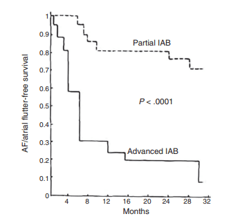
IAB is strongly associated with adverse clinical outcomes. In middle-aged patients, advanced IAB triples the risk of developing atrial fibrillation (AF) and nearly doubles the risk of stroke. It is also associated with increased cardiovascular mortality and sudden death, and in older adults it is also associated with all-cause mortality. The relevance of advanced IAB extends to neurological health: its presence shows a progressive association with cognitive decline and dementia, similar to AF. This relationship is probably mediated by silent strokes and/or chronic cerebral hypoperfusion. Because of these findings, it is recommended that patients with advanced IAB undergo routine cognitive screening and that IAB be investigated in cases of cognitive impairment.
Conclusion
Bayés syndrome establishes IAB as a high-risk predictor reflecting an underlying fibrotic atrial cardiomyopathy. The presence of advanced IAB on the ECG is linked to an increased incidence of AF, stroke and dementia. Therefore, this finding is not just an electrocardiographic diagnosis, but an essential warning sign of a patient who requires closer clinical surveillance and consideration of screening for thromboembolic and cognitive complications.
References
- Scheffer MK, De Marchi MFN, de Alencar Neto JN, Felicioni SP. Eletrocardiograma de A a Z. São Paulo: Manole, 2024.
- Bayés de Luna A, Fiol-Sala M, Martínez-Sellés M, Baranchuk A. Current ECG aspects of interatrial block. Hearts. 2021;2(3):419-32.
- Bayés de Luna A, Martínez-Sellés M, Bayés-Genís A, Elosua R, Baranchuk A. What every clinician should know about Bayés syndrome. Rev Española Cardiol (English ed.). 2020;73(9):758-62.
- Bayés de Luna A, Platonov P, Cosio FG, Cygankiewicz I, Pastore C, Baranowski R, et al. Interatrial blocks: a separate entity from left atrial enlargement: a consensus report. J Electrocardiol. 2012;45(5):445-51
- Conde D, Baranchuk A, Bayés De Luna A. Advanced interatrial block as a substrate of supraventricular tachyarrhythmias: A well recognized syndrome. J Electrocardiol. 2015;48(2):135-40.
- Bayés de Luna A, Oter MCR, Guindo J. Interatrial conduction block with retrograde activation of the left atrium and paroxysmal supraventricular tachyarrhythmias: influence of preventive antiarrhythmic treatment. Int J Cardiol. 1989;22(2):147-50.
- Martínez-Sellés M, Elosua R, Ibarrola M, De Andrés M, Díez-Villanueva P, Bayés-Genis A, et al. Advanced interatrial block and P-wave duration are associated with atrial fibrillation and stroke in older adults with heart disease: the Bayes registry. Europace. 2020;22(7):1001-8.