Atrial flutter is a supraventricular arrhythmia with a characteristic electrocardiographic pattern, yet it still confuses many students and even professionals. This article will help you master the diagnosis of flutter by understanding its mechanism, its manifestations on the tracing, and the particularities of atrioventricular (AV) conduction.
🧠 Mechanism of Atrial Flutter
Flutter is a macro-reentrant arrhythmia of the atria. Its most common form is typical flutter, which circulates counterclockwise around the tricuspid annulus in the right atrium. This circuit depends on a slow-conduction region called the cavotricuspid isthmus, between the inferior vena cava and the tricuspid valve annulus.
The atrial activation rate in flutter usually ranges from 240 to 340 bpm. Because the AV node cannot conduct all of these impulses to the ventricles, partial block occurs, most commonly 2:1, but it may be 3:1, 4:1, or variable.
🔁 The F wave: the “sawtooth” on the ECG
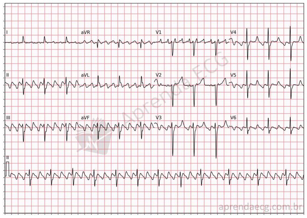
The classic pattern of atrial flutter is the sawtooth F wave, also called “sawtooth waves”, most evident in leads II, III, and aVF. Unlike atrial fibrillation, in which atrial activity is disorganized, flutter shows regular atrial activity, although P waves are absent and replaced by F waves.
The morphology of these waves may vary depending on the direction of the reentrant circuit:
- Typical counterclockwise flutter (most common): negative F waves in II, III, and aVF and positive in V1.
- Typical clockwise flutter: positive F waves in II, III, and aVF and negative in V1.

📉 ECG Criteria
To identify atrial flutter on the ECG, look for:
- Sawtooth F waves, especially visible in II, III, and aVF
- Absence of an isoelectric baseline between F waves
- Atrial rate between 240 and 340 bpm
- Regular or irregular ventricular rhythm, depending on AV conduction
- Usually narrow QRS complexes (except in cases with aberrant conduction)
🔄 AV conduction can be irregular
Although classic flutter is often associated with a regular ventricular rhythm with fixed AV block (such as 2:1), flutter can also present with variable conduction through the AV node. This matters, because many people mistake this pattern for atrial fibrillation.
What causes the irregularity?
Variable AV conduction may occur spontaneously or be induced by drugs that alter AV nodal refractoriness (such as beta-blockers, calcium channel blockers, or digoxin). In these situations, the RR interval becomes irregular, but atrial activity remains regular: this is the key to the correct diagnosis.