The Brugada algorithm, developed in 1991, introduced an innovative approach to the differential diagnosis between ventricular tachycardia (VT) and supraventricular tachycardia with aberrant conduction (SVT-A). It was the first method to offer a sequential framework for distinguishing these complex conditions and has since become one of the most widely used tools in the diagnosis of wide QRS tachycardias.
Introduction to the Brugada Algorithm
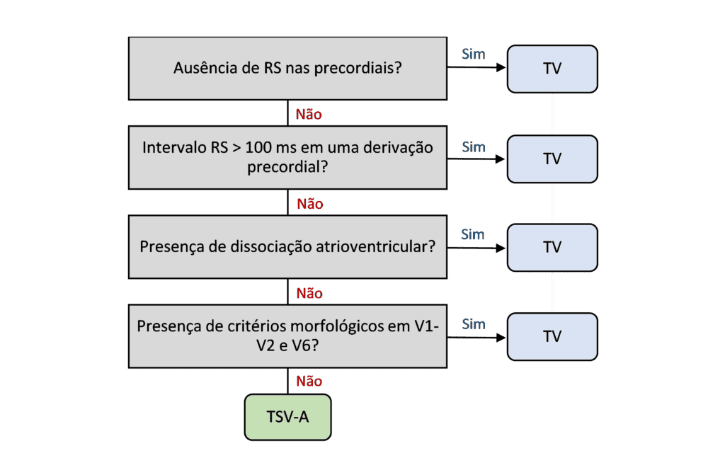
The Brugada algorithm consists of four sequential steps, each with well-defined criteria that make it easier to interpret the electrocardiogram (ECG) during episodes of wide QRS tachycardia. Applying these steps makes it possible to determine whether the tachycardia originates in the ventricles (VT) or is a supraventricular tachycardia with aberrant conduction (SVT-A). If any step yields a positive answer, VT is suggested; if all answers are negative, SVT-A is suggested.
Steps of the Brugada Algorithm
- Presence of an RS complex in all precordial leads: if no RS complex is visible in any of the precordial leads, the diagnosis of VT is confirmed, since the absence of an RS complex is highly specific for VT.
- RS interval longer than 100 ms: if an RS complex is present, the interval from the onset of the R wave to the nadir of the S wave is measured. If it exceeds 100 ms, VT is diagnosed. This criterion is based on the fact that initial ventricular activation is usually slower in VT than in SVT-A.
- Atrioventricular dissociation: the presence of AV dissociation is specific for VT, indicating that the atria and ventricles are out of sync, which is characteristic of a ventricular origin.
- Morphological criteria: finally, if the diagnosis has not yet been reached, QRS morphology is analyzed in V1, V2 and V6.
Morphological Criteria
- Leads with a right bundle branch block (RBBB) pattern:
- In V1: a monophasic R wave or a QR complex indicates VT.
- In V6: a QS or QR complex suggests VT, especially when an RBBB pattern is present.
- Leads with a left bundle branch block (LBBB) pattern:
- In V1 and V2: a broad R wave (longer than 30 ms) or a notched downstroke of the S wave (notch sign) is characteristic of VT.
- In V6: a QS or QR complex favors the diagnosis of VT.
Conclusion
The Brugada algorithm revolutionized the diagnostic approach to wide QRS tachycardias and is a practical and reliable tool for distinguishing VT from SVT-A. It is essential for physicians and students of cardiology and electrophysiology, providing a solid basis for fast and accurate ECG interpretation in emergency settings.
To explore this topic further and deepen your knowledge of tachycardia diagnosis and other ECG topics, check out the ECG library at Aprenda ECG.
Read Also
References
- Scheffer MK, De Marchi MFN, de Alencar Neto JN, Felicioni SP. Eletrocardiograma de A a Z. São Paulo: Manole, 2024.
- Brugada P, Brugada J, Mont L, Smeets J, Andries EW. A new approach to the differential diagnosis of a regular tachycardia with a wide QRS complex. Circulation. 1991 May;83(5):1649–59.