Wide QRS complex tachycardias are a challenging and critical condition to manage in the emergency setting. Studies show that 80% of these tachycardias are ventricular tachycardias (VT), a figure that rises to 90% in the presence of underlying structural heart disease. In this article, we explore the key points in managing these arrhythmias, highlighting the most recent recommendations and the importance of not relying exclusively on electrocardiographic differentiation criteria.
Criteria to Differentiate VT from SVT-A
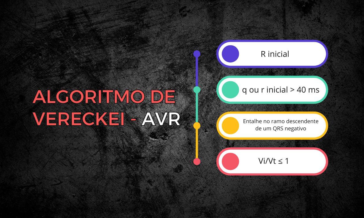
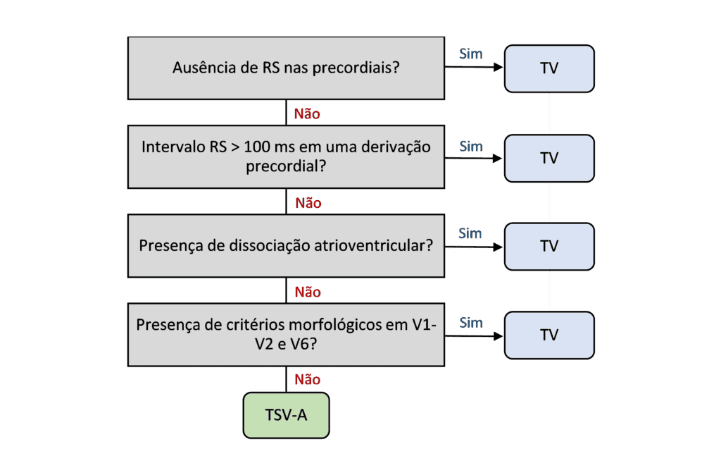
Although there are criteria, such as those of Brugada and Vereckei, to differentiate ventricular tachycardia (VT) from supraventricular tachycardia with aberrancy (SVT-A), these criteria have limitations in emergency clinical practice. The negative likelihood ratio of the Vereckei criteria is 0.34, while that of the Brugada criteria is 0.24. This means that even when they are applied, there is a considerable post-test probability (68-75%) of VT, even with negative criteria.
Another complicating factor is the lack of baseline electrocardiograms (ECGs) for patients. Patients with congenital heart disease or bundle branch blocks may show ECG patterns that mimic ventricular tachycardia, leading to misinterpretation. In addition, idiopathic VT, such as fascicular VT, often presents with patterns that mimic bundle branch block, increasing the risk of diagnostic confusion.
Focus on the Patient's Clinical Status
When facing a patient with a wide QRS complex tachycardia, the most important thing is to assess the patient's clinical status. If there are signs of clinical deterioration, such as chest pain, dyspnea or signs of shock, immediate synchronized electrical cardioversion is indicated, according to both American and European guidelines. In some cases, applying algorithms can distract less experienced healthcare professionals, shifting the focus from urgent clinical treatment to diagnostic analyses that can be done later.
When the patient is hemodynamically stable, adenosine can be used as a therapeutic test, since it can terminate some SVT-A and even some forms of VT. If adenosine is not effective, intravenous antiarrhythmics such as procainamide or amiodarone are recommended. It is important to note that the European guidelines differ slightly, recommending electrical cardioversion in hemodynamically stable patients with wide QRS tachycardia, provided that the anesthetic risk is low.
Post-Stabilization Considerations
After the patient is stabilized, involvement of a cardiologist or electrophysiologist is essential to determine the definitive treatment plan. The electrocardiogram, together with complementary tests such as echocardiography or cardiac magnetic resonance imaging, can provide important information about the etiology of the tachycardia and help in choosing between treatments such as ablation or defibrillator implantation.
Conclusion
Although electrocardiographic criteria for differentiating VT from SVT-A are valuable tools, they should not be the main focus in the acute management of wide QRS tachycardias in unstable patients. Immediate clinical management should prioritize stabilizing the patient, with a focus on electrical cardioversion in cases of clinical deterioration. Algorithms may be more appropriate in later phases, after stabilization, to guide definitive treatment.
References
- Alencar JN De, Carvalho GD De, Campelo RT, Felicioni SP, Scheffer MK, Marchi MN De. Manejo de Taquicardias de Complexo QRS Alargado na Sala de Emergência: O Que Realmente Importa. Arq Bras Cardiol. 2024;121(6):1–4.