

Ventricular tachycardia (VT) is a potentially serious arrhythmia arising from the ventricles, defined by the presence of three or more consecutive ventricular complexes at a HR > 100 bpm.
Electrocardiographic Criteria
Monomorphic ventricular tachycardia
- Regular wide QRS tachycardia
- Electrocardiographic signs suggestive of VT: AV dissociation, morphological criteria, extreme QRS axis deviation, positive or negative precordial concordance, RS interval > 100 ms in precordial leads with RS morphology, R-wave peak time in lead II ≥ 50 ms, initial R wave in aVR, etc.
Polymorphic ventricular tachycardia
- Multiple ventricular foci resulting in QRS complexes that change morphology from beat to beat, in amplitude, axis, and/or duration
- Usually nonsustained and irregular
Differential Diagnosis of Wide QRS Tachycardias
- There are several causes of so-called wide QRS tachycardias, including VT. Diagnostic algorithms help us differentiate VT from SVT with aberrant conduction
- The main algorithms include Brugada, Vereckei-aVR, Santos, Basel, etc.
- See the new page on Wide QRS Tachycardias for more information about the algorithms
Classification of Ventricular Tachycardias
By duration:
- Nonsustained (NSVT): ends spontaneously within 30 seconds
- Sustained: lasts > 30 s and/or causes hemodynamic instability
By morphology:
- Monomorphic: a single QRS complex morphology
- Polymorphic: more than one QRS complex morphology
By hemodynamic stability:
- Stable: no hemodynamic compromise
- Unstable: hemodynamic compromise, manifested by chest pain, dyspnea, hypotension, and/or syncope
ECG in Ventricular Tachycardia
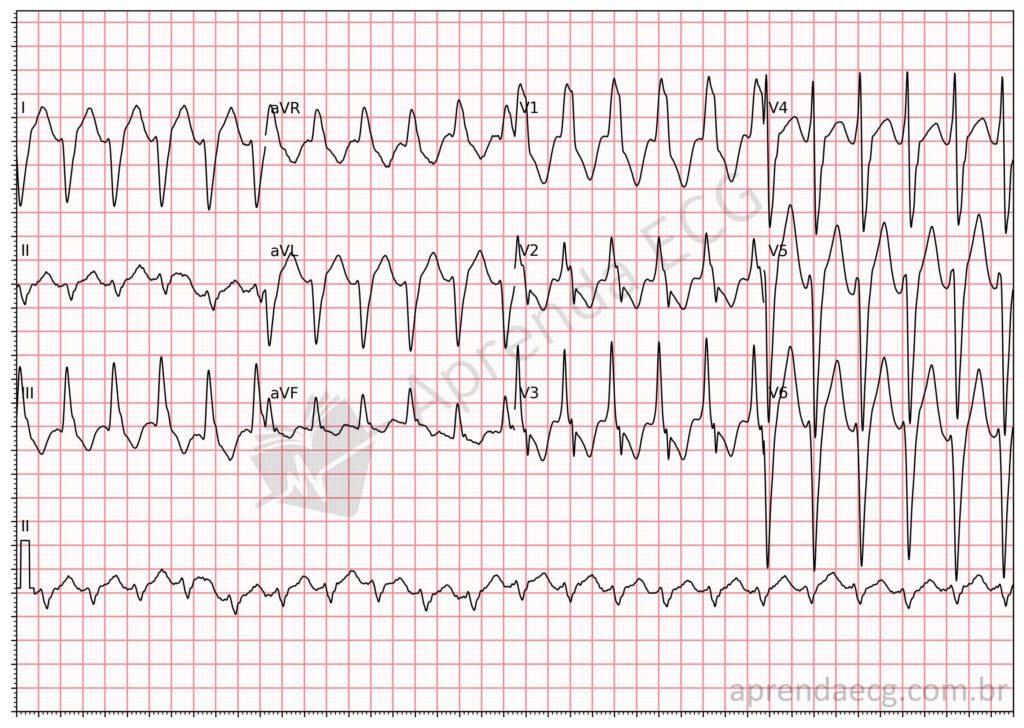
Wide QRS tachycardia suggestive of ventricular tachycardia. ECG showing multiple electrocardiographic criteria suggestive of VT. Try applying the Brugada and Vereckei algorithms.
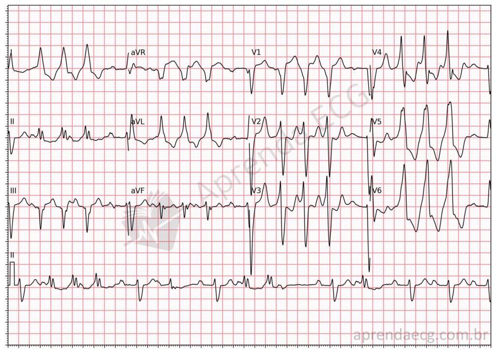
Nonsustained ventricular tachycardia. NSVT consists of a run of 3 or more beats of ventricular origin lasting less than 30 seconds and without hemodynamic instability. In this example, the first beat is sinus, followed by 3 beats of ventricular origin. Next, isolated and paired PVCs are also seen. The underlying rhythm is sinus with first-degree AV block and a widened QRS.
Read Also
Related Topics
References
- Scheffer MK, De Marchi MFN, de Alencar Neto JN, Felicioni SP. Eletrocardiograma de A a Z. São Paulo: Manole, 2024.
- Alencar JN De, Carvalho GD De, Campelo RT, Felicioni SP, et al. Manejo de Taquicardias de Complexo QRS Alargado na Sala de Emergência: O Que Realmente Importa. Arq Bras Cardiol. 2024;121(6):1–4.
- Alencar Neto JN de. Tratado de ECG. Salvador: Sanar; 2022. 816 p.
- Zeppenfeld K, Tfelt-Hansen J, de Riva M, et al. 2022 ESC Guidelines for the management of patients with ventricular arrhythmias and the prevention of sudden cardiac death Developed by the task force for the management of patients with death of the European Society of Cardiology ( ESC ). Eur Heart J. 2022;1–130.


