
Wide QRS tachycardia, with QRS complexes longer than 120 ms and a heart rate above 100 bpm, is usually caused by ventricular tachycardia (VT) or supraventricular tachycardia with aberrancy (SVT-A). Several diagnostic algorithms, such as the Brugada algorithm and the Vereckei aVR algorithm, have been developed to help differentiate between these two conditions with greater diagnostic precision. Although algorithms increase the accuracy of the assessment, the diagnosis must be complemented by clinical experience and careful analysis of the electrocardiogram.
Causes of Wide QRS Tachycardia
The causes of wide QRS tachycardia are diverse, encompassing both ventricular arrhythmias and supraventricular conditions with altered conduction mechanisms. The main causes include:
- Ventricular tachycardia (VT): one of the most common causes, characterized by its origin in the ventricles and frequently associated with structural heart disease.
- Supraventricular tachycardia with aberrant conduction: occurs when a supraventricular tachycardia (SVT) is conducted with aberrancy due to a preexisting or functional bundle branch block, which increases QRS duration.
- Supraventricular tachycardia with conduction over an accessory pathway: in this case, the wide QRS may be caused by preexcited SVT or by antidromic atrioventricular reentrant tachycardia (AVRT), in which activation travels over an accessory pathway.
- Supraventricular tachycardia with antiarrhythmic drugs: some class IA and IC antiarrhythmic drugs can prolong QRS duration, producing a wide QRS pattern even in supraventricular arrhythmias.
- Electrolyte disturbance (hyperkalemia): severe hyperkalemia can alter cardiac electrical conduction, resulting in a wide QRS pattern.
- Ventricular-paced rhythm: in patients with a pacemaker, direct ventricular pacing also produces a wide QRS, since the electrical impulse does not follow the heart's natural conduction system.
Wide QRS Tachycardia Algorithms
Below we present the Brugada, Vereckei, and Basel algorithms, widely used to aid in the differential diagnosis of wide QRS tachycardias. These algorithms provide specific criteria to distinguish ventricular tachycardias from supraventricular tachycardias with aberrant conduction, enabling a systematic approach and improving diagnostic accuracy.
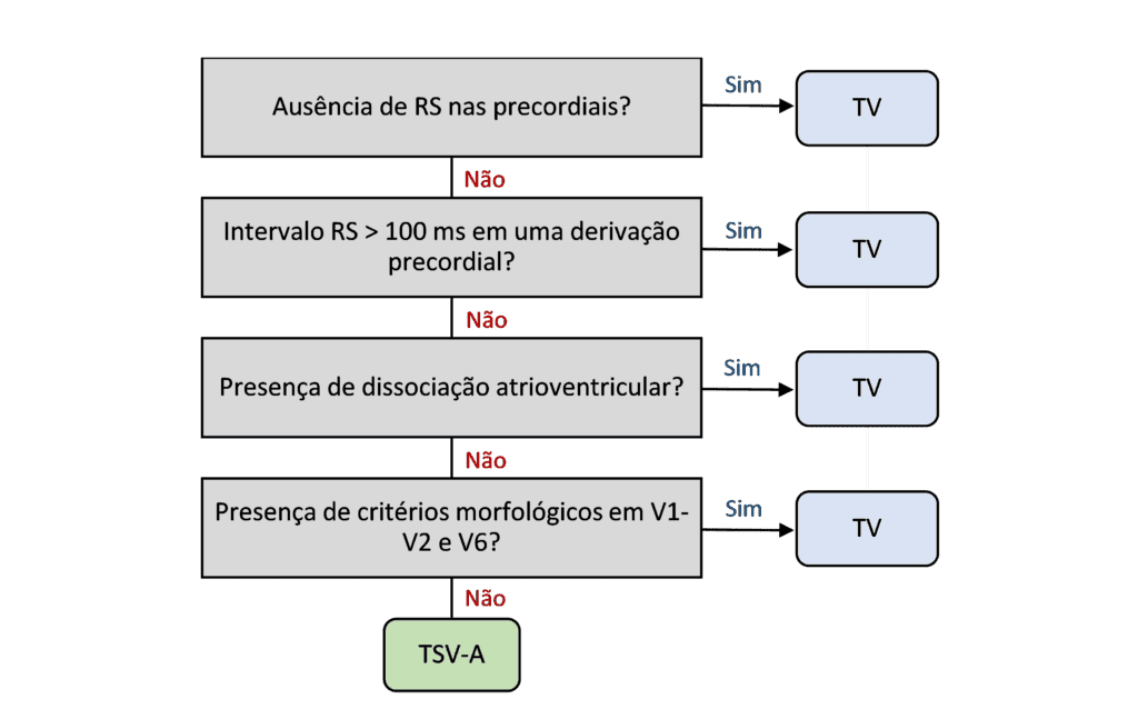
Brugada Algorithm
Published in 1991 by brothers Josep and Pedro Brugada, the Brugada algorithm was the first systematic algorithm created to help differentiate ventricular tachycardia (VT) from supraventricular tachycardia with aberrancy (SVT-A).
Based on a logical sequence of four steps, the algorithm analyzes specific ECG features. If any of the criteria is positive, the diagnosis favors ventricular tachycardia.
Even with the emergence of newer algorithms, the Brugada algorithm remains the best known, most widely taught, and most used worldwide, and it is a landmark in the approach to wide QRS tachycardias.
Vereckei aVR Algorithm
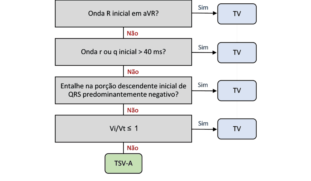
The Vereckei aVR algorithm was published in 2008 by László Vereckei and colleagues as a simplified alternative for differentiating wide QRS tachycardias and has become a useful tool because of its simplicity.
What sets this algorithm apart is its exclusive focus on lead aVR, allowing a faster, more practical analysis, especially useful in emergencies or when the tracing is difficult to interpret.
Like the Brugada algorithm, it consists of a sequence of up to four steps. If any of the criteria is positive, the diagnosis favors ventricular tachycardia.
Basel Algorithm
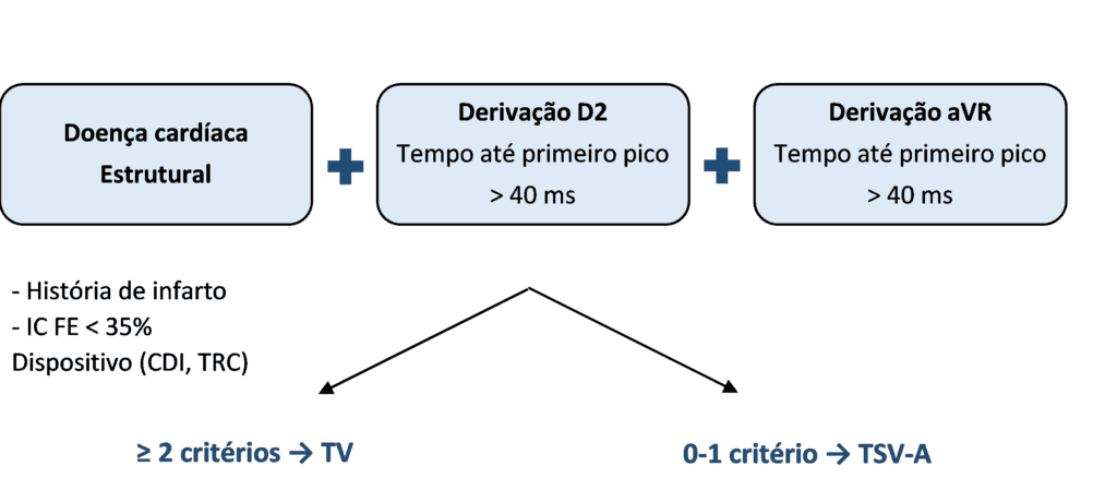
The Basel algorithm, described in 2022 by Moccetti and colleagues from the University of Basel, Switzerland, proposed an integrated approach combining clinical and electrocardiographic criteria to differentiate ventricular tachycardia (VT) from supraventricular tachycardia with aberrancy (SVT-A).
Its logic is simple and effective: VT is diagnosed if at least 2 of the following 3 criteria are present:
- Clinical criterion: a “high-likelihood” finding, such as a history of structural heart disease.
- ECG criterion, lead II: time from QRS onset to the first peak > 40 ms.
- ECG criterion, lead aVR: time to the first peak > 40 ms.
References
- Scheffer MK, De Marchi MFN, de Alencar Neto JN, Felicioni SP. Eletrocardiograma de A a Z. São Paulo: Manole, 2024.
- Alencar JN De, Carvalho GD De, Campelo RT, Felicioni SP, et al. Manejo de Taquicardias de Complexo QRS Alargado na Sala de Emergência: O Que Realmente Importa. Arq Bras Cardiol. 2024;121(6):1–4.
- Brugada P, Brugada J, Mont L, Smeets J, et al. A new approach to the differential diagnosis of a regular tachycardia with a wide QRS complex. Circulation. 1991;83(5):1649–59.
- Vereckei A, Duray G, Szénási G, Altemose GT, et al. New algorithm using only lead aVR for differential diagnosis of wide QRS complex tachycardia. Hear Rhythm. 2008;5(1):89–98.
- Moccetti F, Yadava M, Latifi Y, Strebel I, et al. Simplified Integrated Clinical and Electrocardiographic Algorithm for Differentiation of Wide QRS-Complex Tachycardia. JACC Clin Electrophysiol. 2022.