Matheus Kiszka Scheffer, MD
Introduction
On August 28, 2026, during the ESC Congress, the European Society of Cardiology, the American College of Cardiology, the American Heart Association, and the World Heart Federation jointly published the Fifth Universal Definition of Myocardial Infarction (5th UDMI), eight years after the fourth version.1,2 The document reorganizes the etiologic classification of myocardial infarction, makes the sex-specific 99th percentile of troponin mandatory, and devotes an entire section to the electrocardiogram.1
For ECG readers, the changes lie less in the ST-elevation thresholds, which remained the same, and more in three points: the explicit recognition that some infarctions without ST elevation have an occluded culprit artery, the list of patterns that can be treated as STEMI equivalents, and the return to a single definition of the pathologic Q wave.1 This article summarizes what has changed, compares the criteria with those of the 4th definition3, and points out what remains open.
What changed in the definition
The numbering of types 1 to 5 has been abandoned. Myocardial infarction is now classified into three clinical types according to mechanism:1
- Primary: resulting from acute coronary pathology (atherothrombosis, spontaneous dissection, spasm, or embolism). It includes restenosis, stent thrombosis, and graft failure occurring more than 30 days after revascularization.
- Secondary: the result of an imbalance between oxygen supply and demand caused by another acute condition. For a confirmed diagnosis, the document requires obstructive coronary artery disease (stenosis ≥70%, or ≥50% with flow limitation on physiological testing) without acute coronary pathology, or a new regional wall motion abnormality, or loss of viability on imaging.
- Procedure-related: a coronary complication within 30 days of any cardiac procedure. A confirmed diagnosis now depends on imaging; multiples of troponin above the 99th percentile serve only as a trigger for investigation.
The former type 3, sudden cardiac death before biomarker sampling, no longer exists as a category. When autopsy or postmortem imaging is available, the death is reclassified into one of the three types.1
Myocardial injury continues to be defined by a troponin elevation above the 99th percentile, which must now be sex-specific; with high-sensitivity troponin T, the female limit corresponds to about half of the male limit.1 The acronym MINOCA was kept, with a new meaning: myocardial injury with nonobstructive coronary arteries. The change acknowledges that this is a working diagnosis; magnetic resonance imaging confirms infarction in 22% to 27% of these patients.1
Myocardial infarction still requires acute myocardial injury associated with clinical evidence of acute ischemia. It is in this second component that the ECG comes in.
Electrocardiographic criteria for ischemia
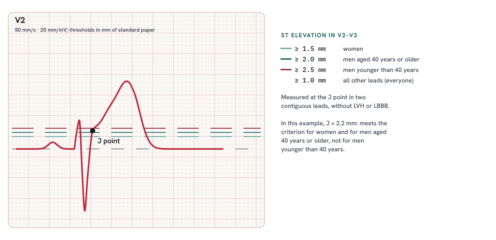
The ST-segment elevation thresholds are the same as in the 4th definition: new ST elevation at the J point in at least two contiguous leads, with specific values for V2–V3, applicable in the absence of left ventricular hypertrophy and left bundle branch block.1,3 The differences appear in T-wave inversion, in the posterior leads, and in the Q wave (Table 1).
| Criterion | 4th UDMI (2018) | 5th UDMI (2026) |
|---|---|---|
| ST elevation in V2–V3 | ≥2 mm in men ≥40 years; ≥2.5 mm in men <40 years; ≥1.5 mm in women | No change |
| ST elevation in other leads | ≥1 mm | No change |
| ST depression | New, horizontal or downsloping, ≥0.5 mm in two contiguous leads | No change |
| T-wave inversion | >1 mm in two contiguous leads with prominent R wave or R/S ratio >1 | ≥1 mm in two contiguous leads, new or dynamic (in Table 5 of the document; the text uses >1 mm); the prominent R wave requirement is not repeated |
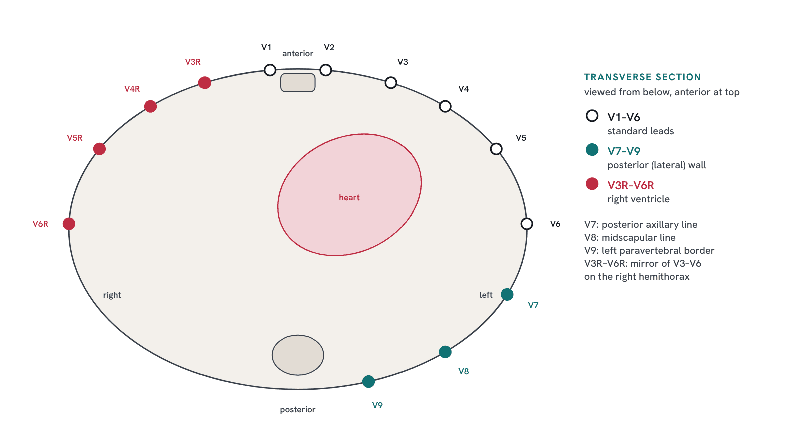
| Posterior (lateral) wall* | ST elevation ≥0.5 mm in V7–V9 (≥1 mm in men <40 years) | ST depression ≥1 mm in V1, V2, and/or V3, especially with a dominant R wave, confirmed by ST elevation in V7–V9, with no numerical threshold for V7–V9 |
| Right ventricle | ST elevation ≥0.5 mm in V3R–V4R (≥1 mm in men <30 years) | ST elevation in V3R–V6R, especially when accompanied by ST elevation in aVR, with no numerical threshold |
| Pathologic Q wave | Different criteria by territory (>20 ms in V2–V3; ≥30 ms and ≥1 mm in the other leads) | Single definition: ≥40 ms and/or depth ≥25% of the R wave, in two contiguous leads |
All findings of ischemia or infarction listed by the 5th UDMI
In addition to the ST-deviation thresholds, Table 5 of the 5th UDMI brings together the other electrocardiographic findings of ischemia or infarction (Table 2).1
| Finding | Description in the 5th UDMI |
|---|---|
| ST elevation | New ST elevation at the J point in two contiguous leads: ≥1 mm in all leads except V2–V3, where the thresholds are ≥2.5 mm in men <40 years, ≥2 mm in men ≥40 years, and ≥1.5 mm in women of any age, in the absence of bundle branch block and/or left ventricular hypertrophy |
| ST depression | New horizontal or downsloping ST depression ≥0.5 mm at the J point in two or more contiguous leads |
| Hyperacute T wave | Symmetric, broad-based T waves, disproportionately large relative to the preceding QRS, in two contiguous leads |
| de Winter T wave | Tall, prominent, symmetric T waves with upsloping ST depression in the precordial leads |
| Biphasic T wave | Initial positive deflection followed by a negative deflection |
| T-wave inversion | New or dynamic inversion ≥1 mm in two contiguous leads |
| Wellens syndrome | Biphasic or deeply inverted T waves in V2 and V3 |
| Pathologic Q wave | Duration ≥40 ms and/or depth ≥25% of the R wave in two contiguous leads |
| Sgarbossa criteria | In the presence of left bundle branch block or ventricular pacing: concordant ST elevation ≥1 mm; ST depression ≥1 mm in V1, V2, or V3; discordant ST elevation ≥5 mm |
| Ventricular arrhythmia | Ventricular fibrillation or ventricular tachycardia |
Three items in the table broaden the traditional reading centered on the ST segment. The hyperacute T wave receives its own definition: symmetric, broad-based, and disproportionate to the QRS that precedes it. The biphasic T wave, with an initial positive and a terminal negative component, appears as an isolated finding, in addition to being part of Wellens syndrome. And ventricular arrhythmias, ventricular fibrillation or tachycardia, are now listed among the electrocardiographic manifestations of acute ischemia.1
The document also describes the patient who presents late after a coronary occlusion: in this setting, pathologic Q waves and biphasic or inverted T waves predominate, rather than ST deviation, although some of these patients never develop a Q wave.1 There is a small internal inconsistency in the T-wave inversion threshold, which appears as ≥1 mm in Table 5 and as >1 mm in the text; in practice, the finding must be new or dynamic and present in two contiguous leads.1
The document also lists causes of nonischemic ST elevation that should be considered before attributing the finding to infarction: early repolarization, pericarditis, myocarditis, Brugada syndrome, Takotsubo syndrome, pulmonary embolism, hyperkalemia, hypothermia, and intracranial hypertension. In coronary occlusion, ST elevation is usually regional and accompanied by reciprocal ST depression; in pericarditis and early repolarization, it tends to be diffuse, not confined to a coronary territory, and without reciprocal changes.1
Acute coronary occlusion without ST elevation
The 5th UDMI states that up to one in four patients managed as NSTEMI has an occluded culprit artery.1 This figure is consistent with the meta-analysis by Khan et al., which pooled 40,777 patients and found culprit artery occlusion in 25.5% of NSTEMIs, with higher short-term mortality in this group (relative risk 1.67).4
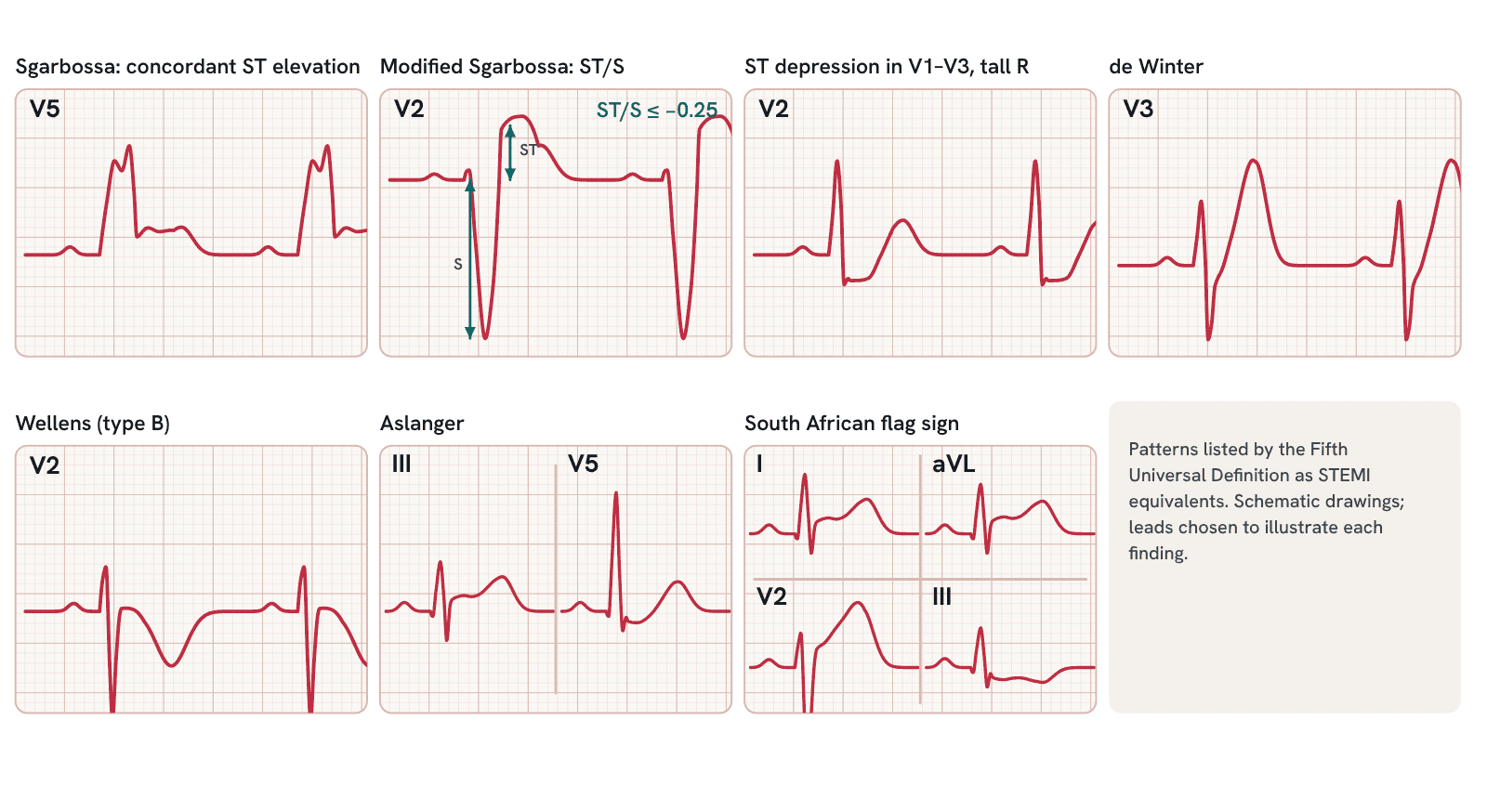
To narrow this gap, the document lists patterns that may be considered STEMI equivalents:1
- new or presumed new left or right bundle branch block, accompanied by other signs of ischemia;
- Sgarbossa criteria (concordant ST elevation ≥1 mm; ST depression ≥1 mm in V1, V2, or V3; discordant ST elevation ≥5 mm) and the modified Sgarbossa criterion (ST/S ratio ≤0.25), also applicable to ventricular paced rhythm;
- marked ST depression or hyperacute T waves in V1–V2, with reciprocal changes in other leads;
- de Winter pattern (upsloping ST depression with tall, symmetric T waves in V2–V5) and Wellens pattern (biphasic or deeply inverted T waves in V2–V3 during pain-free intervals);
- Aslanger pattern: isolated ST elevation in lead III with ST depression in any lead from V4 to V6 and a positive T wave;
- South African flag pattern: ST elevation in leads I, aVL, and V2 with ST depression in lead III.
The text also allows a diagnosis of STEMI in the presence of "other changes suggestive of acute coronary occlusion."1 In practice, the definition moves closer to the occlusion MI paradigm (OMI/NOMI), proposed by Stephen Smith and Pendell Meyers, which shifts the focus from ST elevation to the presence of acute occlusion.5 In a case-control study of 808 patients, the classic STEMI criteria identified 41% of occlusions, compared with 86% when the ECG was read by an interpreter trained in OMI findings, with similar specificity (94% and 91%).6
The document, however, does not adopt the OMI/NOMI nomenclature. It refers to "acute coronary occlusion" and keeps the STEMI/NSTEMI dichotomy, including in the ICD-11 codes.1 For the ECG reader, the practical consequence is the same: the absence of ST elevation meeting the thresholds does not exclude occlusion, and the patterns listed above warrant immediate discussion with the cardiac catheterization team.
Q waves, unrecognized infarction, and additional leads
The pathologic Q wave once again has a single definition: duration ≥40 ms and/or depth ≥25% of the R wave, in two contiguous leads.1 The document justifies this choice by its better correlation with transmural infarction on magnetic resonance imaging and abandons the territory-specific criteria of the previous version.1,3
At the same time, the 5th UDMI acknowledges that an isolated Q wave has limited specificity for unrecognized infarction and recommends confirmation by imaging, preferably cardiac magnetic resonance with late gadolinium enhancement.1 Silent infarction is common: in studies with systematic magnetic resonance imaging, it appears in about one in five people over 70 years of age and in 3% of asymptomatic individuals with a mean age of 50 years.1
For the lateral wall*, the document describes ST depression ≥1 mm in V1, V2, and/or V3, especially when the R wave is larger than the S wave (dominant) in V1 or V2, as a sign to be confirmed by ST elevation in leads V7 to V9. For the right ventricle, it cites ST elevation in V3R to V6R, especially when accompanied by ST elevation in aVR. In neither case is there a numerical threshold for the additional leads; the values of 0.5 mm and 1 mm that circulate in lectures and textbooks come from the 4th definition.1,3
*Readers of Aprenda ECG know that we prefer the term lateral wall. A true posterior wall practically does not exist: in most people, the basal segment of the inferior wall follows the same plane as the other segments of that wall, without curving upward, and the region formerly called posterior corresponds to the inferobasal segment.7 The correlation between the ECG and contrast-enhanced magnetic resonance imaging showed that a prominent R wave in V1 results from infarction of the lateral wall, and the committee of the International Society for Holter and Noninvasive Electrocardiology proposed abandoning the term posterior.7,8 We kept the word posterior at its first mention only because it is the term used by the 5th UDMI.
Diffuse ST depression with ST elevation in aVR is mentioned as a possible marker of left main or three-vessel disease, and hyperkalemia appears among the causes of nonischemic ST elevation.1 In settings without troponin or imaging, the definition allows the diagnosis to be based on symptoms associated with a new ischemic change or a new Q wave on the ECG, which reinforces the role of the test in low-resource settings.1
What remains open
In 2020, the International Society of Electrocardiology and the International Society for Holter and Noninvasive Electrocardiology published a counterpoint paper on the electrocardiographic part of the 4th definition, with suggestions for the next version.9 Some were incorporated: the de Winter pattern appears by name, and caution regarding diffuse ST depression in left ventricular hypertrophy and left bundle branch block is present.1,9
Other proposals do not appear in the text of the 5th UDMI. Among them: treating an inverted T wave with an isoelectric ST segment as a post-ischemic change rather than active ischemia; adjusting the normal limits of the ST segment by ethnicity; placing V7–V9 on the same horizontal plane as V6; and describing STEMI with spontaneous reperfusion as a distinct entity.9 These points still depend on the judgment of the person interpreting the tracing.
Summary
- The Fifth Universal Definition of Myocardial Infarction (2026) classifies myocardial infarction as primary, secondary, or procedure-related; type 3 has been eliminated.
- The 99th percentile of troponin must now be sex-specific.
- The thresholds for ST elevation and ST depression have not changed from 2018.
- New or dynamic T-wave inversion ≥1 mm in two contiguous leads (>1 mm in the text of the document) no longer repeats the prominent R wave requirement.
- The pathologic Q wave has a single definition (≥40 ms and/or ≥25% of the R wave), and an isolated Q wave should be confirmed by imaging.
- The document acknowledges that up to one in four NSTEMIs has an occluded culprit artery and lists STEMI equivalents (Sgarbossa and modified Sgarbossa, de Winter, Wellens, Aslanger, South African flag, new bundle branch block with ischemia).
- The OMI/NOMI nomenclature was not adopted, but a diagnosis of STEMI is allowed in the presence of changes suggestive of acute coronary occlusion.
- Table 5 of the 5th UDMI includes, in addition to ST deviation, the hyperacute T wave, de Winter T wave, biphasic T wave, Wellens syndrome, pathologic Q wave, Sgarbossa criteria, and ventricular arrhythmia. Leads V7–V9 and V3R–V6R are cited without their own numerical threshold, and we prefer the term lateral wall to “posterior” wall.
References
- Mills NL, Newby LK, Zaman S, Almahmeed WA, Bucciarelli-Ducci C, Colvin MM, et al. Fifth Universal Definition of Myocardial Infarction (2026). Eur Heart J. 2026. doi:10.1093/eurheartj/ehag101. Published simultaneously in J Am Coll Cardiol. 2026;88(11):1314-68.
- Mills NL, Newby LK, Zaman S, Almahmeed WA, Bucciarelli-Ducci C, Colvin MM, et al. Fifth Universal Definition of Myocardial Infarction (2026). Glob Heart. 2026;21(1):64. doi:10.5334/gh.1578.
- Thygesen K, Alpert JS, Jaffe AS, Chaitman BR, Bax JJ, Morrow DA, et al. Fourth Universal Definition of Myocardial Infarction (2018). Circulation. 2018;138(20):e618-51. doi:10.1161/CIR.0000000000000617.
- Khan AR, Golwala H, Tripathi A, Bin Abdulhak AA, Bavishi C, Riaz H, et al. Impact of total occlusion of culprit artery in acute non-ST elevation myocardial infarction: a systematic review and meta-analysis. Eur Heart J. 2017;38(41):3082-9. doi:10.1093/eurheartj/ehx418.
- Smith SW, Meyers HP. ST elevation is a poor surrogate for acute coronary occlusion. Let's replace STEMI with occlusion MI (OMI)!! Int J Cardiol. 2024;407:131980. doi:10.1016/j.ijcard.2024.131980.
- Meyers HP, Bracey A, Lee D, Lichtenheld A, Li WJ, Singer DD, et al. Accuracy of OMI ECG findings versus STEMI criteria for diagnosis of acute coronary occlusion myocardial infarction. Int J Cardiol Heart Vasc. 2021;33:100767. doi:10.1016/j.ijcha.2021.100767.
- Bayés de Luna A, Rovai D, Pons Llado G, Gorgels A, Carreras F, Goldwasser D, et al. The end of an electrocardiographic dogma: a prominent R wave in V1 is caused by a lateral not posterior myocardial infarction — new evidence based on contrast-enhanced cardiac magnetic resonance–electrocardiogram correlations. Eur Heart J. 2015;36(16):959-64. doi:10.1093/eurheartj/ehv035.
- Bayés de Luna A, Wagner G, Birnbaum Y, Nikus K, Fiol M, Gorgels A, et al. A new terminology for left ventricular walls and location of myocardial infarcts that present Q wave based on the standard of cardiac magnetic resonance imaging: a statement for healthcare professionals from a committee appointed by the International Society for Holter and Noninvasive Electrocardiography. Circulation. 2006;114(16):1755-60. doi:10.1161/CIRCULATIONAHA.106.624924.
- Birnbaum Y, Fiol M, Nikus K, Garcia Niebla J, Bacharova L, Dubner S, et al. A counterpoint paper: comments on the electrocardiographic part of the 2018 Fourth Universal Definition of Myocardial Infarction endorsed by the International Society of Electrocardiology and the International Society for Holter and Noninvasive Electrocardiology. Ann Noninvasive Electrocardiol. 2020;25(5):e12786. doi:10.1111/anec.12786.