

Brugada syndrome is a channelopathy with autosomal dominant inheritance, incomplete penetrance and variable expressivity, responsible for 4% to 12% of all sudden cardiac deaths and up to 20% of those occurring in patients with structurally normal hearts.
Electrocardiographic Criteria
Type 1 Brugada pattern
- Coved ST-segment elevation of at least 2 mm in one or more right precordial leads (V1 to V3), followed by a concave or straight r’ downslope that crosses the baseline and produces a symmetric inverted T wave
Type 2 Brugada pattern
- r’ ≥ 2 mm, ST segment ≥ 0.5 mm above the baseline, descending with downward convexity, followed by a positive T wave in V2 and variable morphology in V1
ECG in Brugada Syndrome
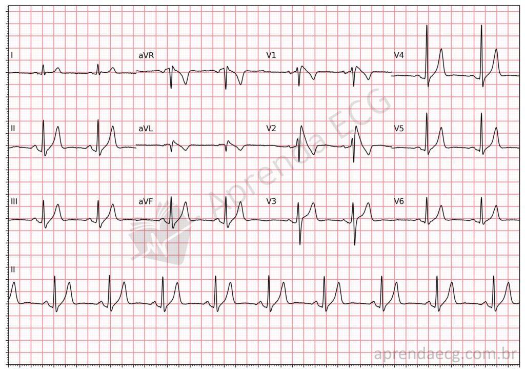
Type 1 Brugada pattern. Note the coved ST-segment elevation in V1 and V2 followed by a straight downslope that crosses the baseline and produces a symmetric inverted T wave.
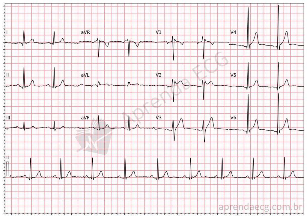
Type 2 Brugada pattern. Note in V2 the r’ wave ≥ 2 mm and ST segment ≥ 0.5 mm, followed by a positive T wave in V2, forming a “saddleback” pattern.
Related Topics
References
- Zeppenfeld K, Tfelt-Hansen J, de Riva M, et al. 2022 ESC Guidelines for the management of patients with ventricular arrhythmias and the prevention of sudden cardiac death Developed by the task force for the management of patients with death of the European Society of Cardiology ( ESC ). Eur Heart J. 2022;1–130.
- Bayés De Luna A, Brugada J, Baranchuk A, Borggrefe M, et al. Current electrocardiographic criteria for diagnosis of Brugada pattern: A consensus report. J Electrocardiol. 2012;45(5):433–42.
- Krahn AD, Behr ER, Hamilton R, Probst V, et al. Brugada Syndrome. JACC Clin Electrophysiol. 2022;8(3):386–405.


