Anterior wall acute myocardial infarction (AMI) carries the highest short-term mortality and the greatest deterioration of ventricular function. The classic electrocardiographic presentation of acute coronary occlusion (OMI) is ST-segment elevation, but this finding is present in only 68% of cases.
With this in mind, a new article published in July 2025 reported the results of a retrospective case-control study conducted by experts, supported by a trained AI, to test how reliably the more traditional criteria can diagnose OMI events in the left anterior descending artery.
What were the results?
The study sample included 808 patients with acute coronary syndrome. Of these 808 participants, 755 were excluded (patients without ECGs in the electronic health record or with insufficient retrospective information to determine the primary outcome, patients with NOMI, that is, no acute coronary occlusion, and patients with OMI of other coronary arteries).
A total of 53 patients with an LAD culprit lesion and TIMI 0 flow (the grade for completely absent blood flow) were analyzed. Of these, 20 patients did not have ST elevation according to current guidelines and definitions (a sensitivity, therefore, of 68%).
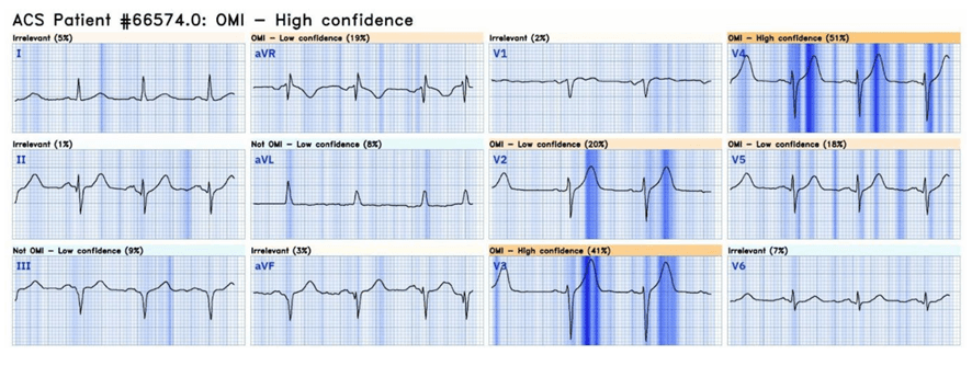
All patients, however, had some other OMI finding not recognized by the guidelines (hyperacute T waves, terminal QRS distortion, etc.; see the table below). In all of these patients, OMI was correctly diagnosed by the human reader familiar with OMI signs and by the Queen of Hearts AI.
Frequency of OMI findings in 20 cases of LAD OMI with TIMI 0 flow that did not meet STEMI criteria
| ECG finding | Frequency | % |
|---|---|---|
| Hyperacute T wave | 17/20 | 85% |
| ST elevation < 1 mm | 17/20 | 85% |
| Pathologic Q wave | 14/20 | 70% |
| Reciprocal changes (ST depression and/or T-wave inversion) | 10/20 | 50% |
| Terminal QRS distortion | 4/20 | 20% |
| Modified Sgarbossa criteria (in LBBB/ventricular paced rhythm) | 0/20 | 0% |
| Maximal ST depression in V2–V4 (posterior) | 0/20 | 0% |
| ST elevation in inferior leads with ST depression/T-wave inversion in aVL | 4/20 | 20% |
AI analysis of the ECG criteria
Patients with OMI without ST elevation had significantly longer door-to-balloon times and higher peak troponin levels (a plausible surrogate for larger infarct size) than patients who were lucky enough to present with ST elevation.
In other words, the OMI/NOMI paradigm and the ECG findings published so far have superior accuracy in diagnosing LAD OMI compared with the “ST elevation” versus “no ST elevation” paradigm.
Summary
ST elevation is present in only 68% of patients with LAD OMI.
The OMI/NOMI paradigm is superior to ST elevation for correctly diagnosing and treating these patients.
References
- Meyers HP, Sharkey SW, Herman R, de Alencar JN, Shroff GR, Frick WH, et al. Failure of standard contemporary ST-elevation myocardial infarction electrocardiogram criteria to reliably identify acute occlusion of the left anterior descending coronary artery. Eur Heart J Acute Cardiovasc Care. 28 de julho de 2025;zuaf037.
- McLaren J, de Alencar JN, Aslanger EK, Meyers HP, Smith SW. From ST-Segment Elevation MI to Occlusion MI: The New Paradigm Shift in Acute Myocardial Infarction. JACC Adv. 2024;3(11):101314.
- Alencar JN De, Feres F, Marchi MFN De, Franchini KG, Scheffer MK, Felicioni SP, et al. Além do Paradigma IAMCSST-IAMSSST: Proposta do Instituto Dante Pazzanese para o Diagnóstico de Oclusão Coronariana Aguda. Arq Bras Cardiol. 2024;121(5):e20230733.
- de Alencar Neto JN, Scheffer MK, Correia BP, Franchini KG, et al. Systematic review and meta-analysis of diagnostic test accuracy of ST-segment elevation for acute coronary occlusion. Int J Cardiol. 2024;131889.
- Este artigo também contou com a valiosa participação de Francisco José Alves da Franca Junior.