
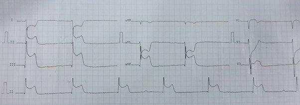
The Dead Man Sign on the ECG is an electrocardiographic curiosity described in inferior acute myocardial infarction (MI), in which the combination of ST-segment elevation and reciprocal changes creates an image on the tracing resembling the silhouette of a man lying down.
It is not a new formal diagnostic criterion, nor does it replace a systematic ECG analysis. Above all, it is an interesting example of how visual memory can help (and sometimes even amuse) those who interpret electrocardiograms.
What Is the Dead Man Sign
The sign was first described as a “new ECG sign” in inferior MI and was later proposed as a possible marker of proximal right coronary artery occlusion.
It occurs in the setting of:
- Inferior ST-elevation MI
- Marked ST elevation in II and aVF (often also in III)
- Reciprocal ST depression in I and aVL
- Frequent association with right ventricular involvement
What is special lies less in the pathophysiology (which is that of classic inferior MI) and more in the geometric pattern formed by the sum of the deflections.
How the Tracing “Draws” the Silhouette
When we look at aVF with marked ST elevation and aVL with reciprocal depression side by side, the curves can form an outline resembling a human body lying supine.
Put simply:
- The convex ST elevation in aVF represents the “torso”
- The transition between the QRS complex and the ST segment gives the impression of a “head”
- The reciprocal depression in aVL suggests the lower outline of the body
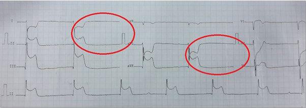
It is a phenomenon of electrocardiographic pareidolia: the brain recognizes familiar patterns in abstract shapes.
Naturally, this depends on the degree of ST elevation and the ST morphology. Not every inferior MI will produce this image.
The Proposed Anatomical Mechanism
In the original reports, the sign was associated with total or near-total occlusion of the proximal right coronary artery.
Some reported features:
- Strong correlation with extensive inferior MI
- Frequent right ventricular involvement
- Association with bradyarrhythmias and tachyarrhythmias (supraventricular or ventricular)
The hypothesis is that a proximal right coronary occlusion produces a stronger inferior injury vector, resulting in exuberant ST elevation in the inferior leads and marked reciprocity in I/aVL.
From an electrophysiological standpoint, therefore, there is no “new mechanism”, only an exuberant graphic manifestation of the inferior injury vector.
Does It Have Practical Value?
Here it is important to separate curiosity from clinical tool.
The diagnosis of inferior MI remains based on the classic criteria of ST-segment elevation in contiguous inferior leads, always together with clinical correlation and hemodynamic risk stratification.
- ST elevation ≥ 1 mm in at least two contiguous inferior leads
- Clinical correlation
- Hemodynamic risk stratification
There is also an often-overlooked technical aspect: the ECG can be displayed in multiple formats. Depending on how the tracing is printed or displayed (sequential by lead, in columns, 3×4, with or without a long rhythm strip, different gain settings), the geometric pattern described may simply not appear. Because the “Dead Man Sign” depends on a specific visual juxtaposition of certain leads, changes in layout can prevent the silhouette from forming, even with the same injury vector.
Its main value is educational:
- It encourages careful morphological observation
- It reinforces the concept of reciprocity
- It helps memorize the pattern of inferior MI with an exuberant vector
In other words: interesting for your visual repertoire. It does not replace systematic analysis.
References
- Tramboo NA, Dar MI, Jahangir, Khan KA, Yaqoob I, Hafeez I. A New ECG Sign. Int J Clin Med Imaging. 2016;3(1).
- Yadav M. Dead Man Sign of ECG- a Unique Predicting Sign of Acute Coronary Syndrome. J Clin Image Case Rep. 2021;5(9):183.