

Left ventricular aneurysm is an area of infarcted myocardium that bulges outward from the ventricular contour during systole and diastole, and it is a possible complication of acute myocardial infarction (MI). It occurs mainly with occlusions of the left anterior descending artery or of a dominant right coronary artery, due to ischemia and the resulting tissue necrosis.
This complication is characterized by pathological changes in left ventricular contractility and myocardial fibrosis, with related electrocardiographic changes, such as electrically inactive areas and secondary changes of the ST-T complex.
Electrocardiographic Criteria
Let's look at the electrocardiographic criteria for ventricular aneurysm:
- ST elevation in at least two contiguous leads
- Q or QS waves in at least two contiguous leads accompanying the ST elevation
- T waves commonly inverted, biphasic, or of low amplitude
- QRS fragmentation may be present
- A prominent R wave in aVR (Goldberger's sign) may be present
Here are some examples:
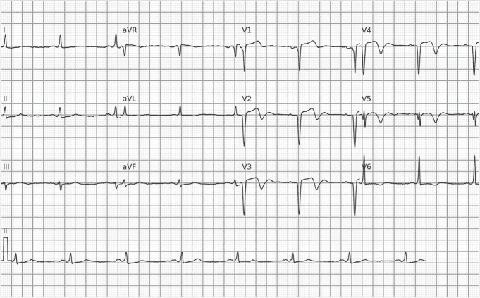
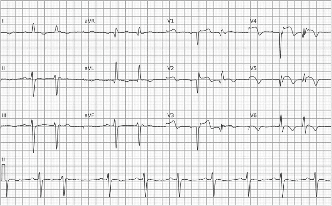
How to Differentiate Ventricular Aneurysm from Anterior Wall OMI
The repolarization changes characteristic of the left ventricular aneurysm pattern make its electrocardiographic differentiation from acute or subacute OMI involving the anterior wall challenging, especially because of the chronic ST elevation and terminal T-wave inversion.
Faced with this challenge, Stephen Smith developed differentiation criteria based on the ratio between the amplitudes of the T wave and the preceding QRS (T/QRS ratio) and on the ratio between their sums from V1 to V4 (ΣT/ΣQRS ratio). These criteria are shown in the table below, with their respective positive (LR+) and negative (LR-) likelihood ratios for OMI:
| Criterion | LR+ | LR- |
|---|---|---|
| T/QRS ≥ 0.36 in at least one lead from V1 to V4 | 4.89 | 0.10 |
| ΣT/ΣQRS ≥ 0.22 (V1 to V4) | 2.93 | 0.12 |
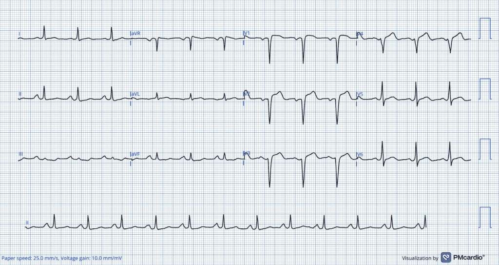
In addition to these criteria, serial electrocardiograms are valuable in differentiating between these two diagnoses. Indeed, a static pattern on serial tracings argues against an acute process, whereas dynamic electrocardiographic changes consistent with an evolving MI favor it. The absence of reciprocal changes also argues against attributing the ST elevation to acute infarction. However, it should be emphasized that the clinical picture is the cornerstone in differentiating the two diagnoses, the ECG being a highly valuable complementary test, but complementary nonetheless.
References
- Smith SW. T/QRS ratio best distinguishes ventricular aneurysm from anterior myocardial infarction. Am J Emerg Med. 2005; 23(3):279–87.
- Klein LR, Shroff GR, Beeman W, Smith SW. Electrocardiographic criteria to differentiate acute anterior ST-elevation myocardial infarction from left ventricular aneurysm. Am J Emerg Med. 2015; 33(6):786–90.
- Scheffer MK, de Alencar Neto JN, De Marchi MFN, Felicioni SP. Eletrocardiograma na síndrome coronária aguda. São Paulo: Manole, 2025.
- Scheffer MK, De Marchi MFN, de Alencar Neto JN, Felicioni SP. Eletrocardiograma de A a Z. São Paulo: Manole, 2024.