
Introduction
Localizing an acute myocardial infarction (AMI) has always been essential for understanding the severity of the event and identifying the culprit artery. For decades, infarct topography was based on pathology studies and correlation with the electrocardiogram (ECG). However, the revolution brought by cardiac magnetic resonance imaging (MRI) redefined this understanding, culminating in a new proposal led by Bayés de Luna. This article reviews that new classification, which today represents the best available evidence on the subject.
Where we came from: the old topographic classification
For much of the 20th century, infarct topography was defined on the basis of autopsies and ECG studies:
- V1-V2: septal wall
- V3-V4: anterior wall
- V5-V6, I, aVL: lateral wall (low and high)
- II, III, aVF: inferior wall
- Tall R in V1-V2: posterior infarction
This classification regarded the “posterior” wall as a region opposite the anterior wall, in contact with the diaphragm. It was Perloff, in the 1960s, who proposed electrocardiographic criteria for “true posterior infarction”. However, this concept began to be questioned as new imaging technologies were developed.
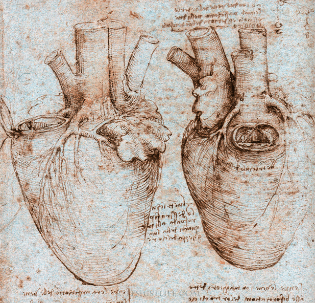
The true orientation of the heart in the chest
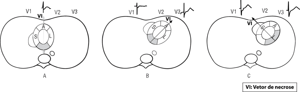
Magnetic resonance imaging showed that the heart lies in an oblique position in the chest, with the left ventricle (LV) facing forward and to the left. This position completely changes our perception of the cardiac walls.
The so-called “posterior wall” is actually the inferobasal segment of the inferior wall, whose electrical activation is late (40–50 ms after QRS onset). Therefore, it cannot cause early R waves in V1-V2. The infarct vector projecting onto these leads actually comes from the lateral wall.
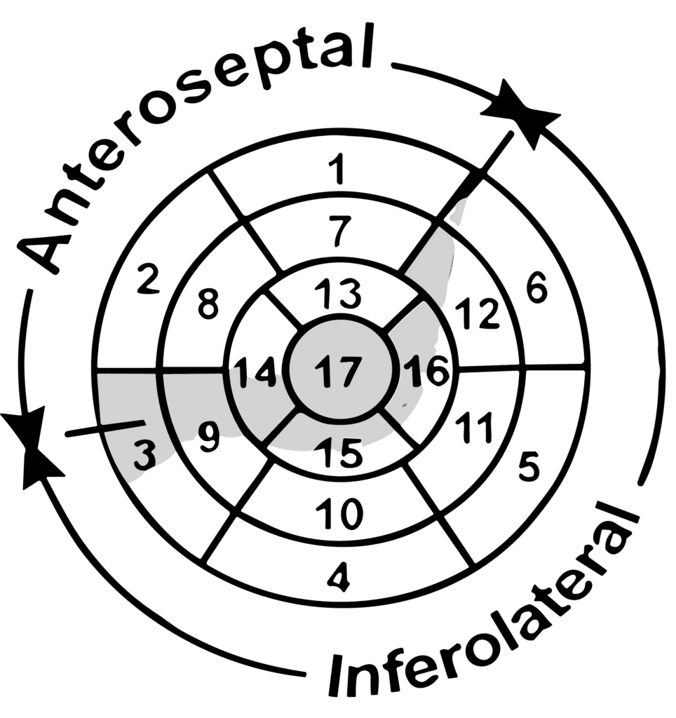
It is also worth noting that the American Heart Association (AHA) classification, published in 2002, helped standardize left ventricular segmentation by proposing a division into 17 segments grouped into four main walls: anterior, septal, lateral, and inferior, with subdivisions into basal, mid, and apical regions and the apex. This model is widely used in imaging studies such as echocardiography, CT, and MRI.
From a functional standpoint, the LV can be divided into two large regions: the anteroapical area, supplied by the left anterior descending artery (LAD), and the inferolateral area, supplied by the right coronary artery (RCA) or the circumflex artery (LCx).
The Bayés de Luna proposal: the best available evidence
In 2006, a group led by Bayés de Luna published in Circulation the new terminology for infarct topography, based on direct correlations between Q-wave patterns (or equivalents) and the areas of fibrosis identified by MRI.
Why is this proposal superior?
- It is based on cardiac MRI, the current gold standard for infarct localization.
- It eliminates confusing terms such as “posterior”, replacing them with more precise locations.
- It has validated sensitivity and specificity for different electrocardiographic patterns.
Infarct Topography Classification According to Bayés de Luna
Below is the table with the new MRI-based classification:
| ECG pattern | Sensitivity and specificity | Name | Most likely culprit artery |
|---|---|---|---|
| Q in V1-V2 | Sens. 100% / Spec. 97% | Septal | Septal branch of the LAD |
| Q from V1-V2 to V3-V6 | Sens. 85% / Spec. 98% | Anteroapical | Mid LAD |
| Q in V1-V2 to V3-V6, I, aVL | Sens. 83% / Spec. 100% | Extensive anterior | Proximal LAD |
| Q in aVL and/or I (sometimes V2-V3) | Sens. 67% / Spec. 100% | Mid-anterior | First diagonal branch |
| Q in I, aVL, V5-V6 and/or RS in V1-V2 | Sens. 67% / Spec. 99% | Lateral | Proximal circumflex artery |
| Q in II, III, aVF | Sens. 88% / Spec. 97% | Inferior | Right coronary or circumflex artery |
| Q in II, III, aVF + Q in I, aVL, V5-V6 and/or RS in V1-V2 | Sens. 73% / Spec. 98% | Inferolateral | Right coronary or circumflex artery |
Clinical impact of the new classification
Understanding the true location of the infarction allows:
- Better correlation with the culprit artery
- More precise interpretation of ECG findings
- Abandonment of ambiguous or incorrect terms (such as “posterior infarction”)
- Alignment with the OMI/NOMI paradigm, which prioritizes detection of acute coronary occlusion based on sensitive electrocardiographic signs
Conclusion
Infarct topography has undergone a true revolution in recent years. Bayés de Luna's MRI-based work today represents the most reliable and best-validated classification for correlating the ECG with the real anatomy of the heart. Adopting this new terminology is essential to improve diagnostic accuracy and clinical reasoning.
References
- Scheffer MK, De Marchi MFN, de Alencar Neto JN, Felicioni SP. Eletrocardiograma de A a Z. São Paulo: Manole, 2024.
- Alencar JN. Tratado de ECG. 2022.
- Bayés de Luna A, Wagner G, Birnbaum Y, Nikus K, Fiol M, Gorgels A, et al. A New Terminology for Left Ventricular Walls and Location of Myocardial Infarcts That Present Q Wave Based on the Standard of Cardiac Magnetic Resonance Imaging. Circulation. 2006;114(16):1755–60.
- Bayés de Luna A. New heart wall terminology and new electrocardiographic classification of Q-wave myocardial infarction based on correlations with magnetic resonance imaging. Rev Esp Cardiol. 2007 Jul;60(7):683–9.
- Cerqueira MD, Weissman NJ, Dilsizian V, Jacobs AK, Kaul S, Laskey WK, et al. Standardized myocardial segmentation and nomenclature for tomographic imaging of the heart. J Cardiovasc Magn Reson. 2002;4(2):203–10.
- Allencherril J, Fakhri Y, Engblom H, Heiberg E, Carlsson M, Dubois-Rande JL, et al. Appropriateness of anteroseptal myocardial infarction nomenclature evaluated by late gadolinium enhancement cardiovascular magnetic resonance imaging. J Electrocardiol. 2018;51(2):218–23.