ST-segment elevation is one of the most recognizable and, at the same time, most complex electrocardiographic findings in modern cardiology. Although it is classic in the setting of ST-elevation myocardial infarction (STEMI), its meaning goes far beyond that, encompassing diagnostic nuances, morphological pitfalls, and relevant prognostic implications. This article aims to review, in a didactic and comprehensive way, everything the cardiologist needs to know about this phenomenon.
🧠 The Origin of the Concept: Harold Pardee's Contributions
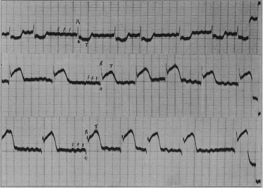
In 1920, Harold Pardee published the first report linking ST-segment elevation to acute obstruction of a coronary artery. The observation was made in a patient with typical anginal symptoms and was confirmed by experimental animal studies, which showed similar changes after ligation of the coronary arteries.
Pardee noted that the abrupt elevation of the ST segment from the J point reflected myocardial injury resulting from transmural ischemia. Following these discoveries, ST elevation became one of the main electrocardiographic indicators of transmural infarction.
📏 How to Measure ST Elevation Correctly
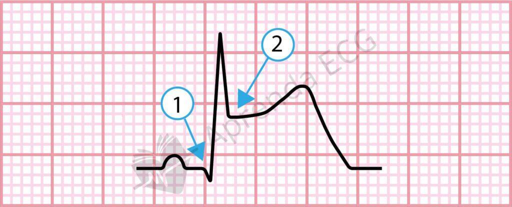
The measurement of ST elevation should be made at the J point (the junction between the QRS and the ST segment) relative to the isoelectric line. The isoelectric line, in turn, can be determined from the TP segment or, preferably, from the PQ junction, especially in patients with tachycardia or an unstable baseline.
Although the cutoffs proposed by the guidelines are useful in clinical practice, especially in population studies and emergency protocols, it is important to remember that they are not absolute. Cases of subtle ST elevation show that relevant ischemic changes can occur even below these thresholds. The table below summarizes these criteria:
| Leads | Men ≥ 40 years | Men < 40 years | Women |
|---|---|---|---|
| V2-V3 | ≥ 2 mm | ≥ 2.5 mm | ≥ 1.5 mm |
| Other leads | ≥ 1 mm | ≥ 1 mm | ≥ 1 mm |
🧐 The Challenge of Subtle ST Elevation
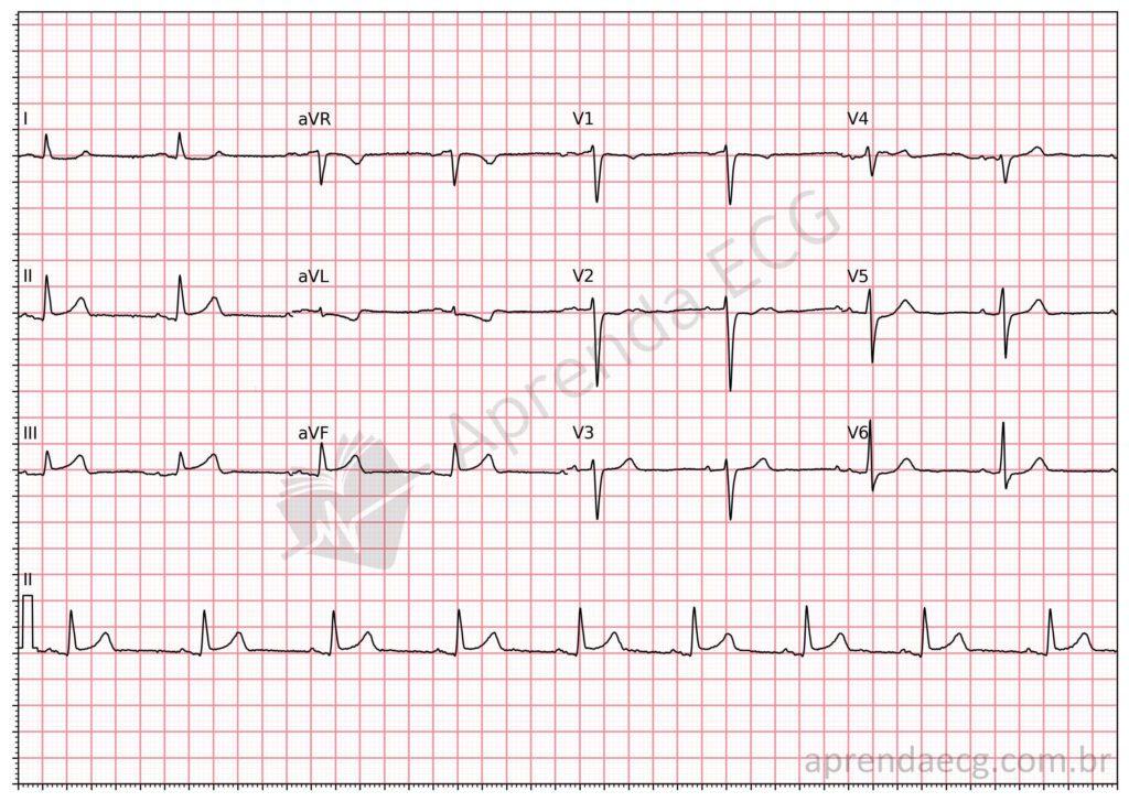
Subtle ST elevation, with elevation between 0.1 mm and 1 mm, is particularly challenging. Studies show that up to 18% of patients with STEMI present with only these subtle changes. This is more common in infarctions involving the circumflex artery (LCx), whose territories generate less activation vector toward the standard leads.
In these cases, the presence of reciprocal ST depression and the clinical assessment become essential.
🔍 ST Morphology: Concave or Convex?
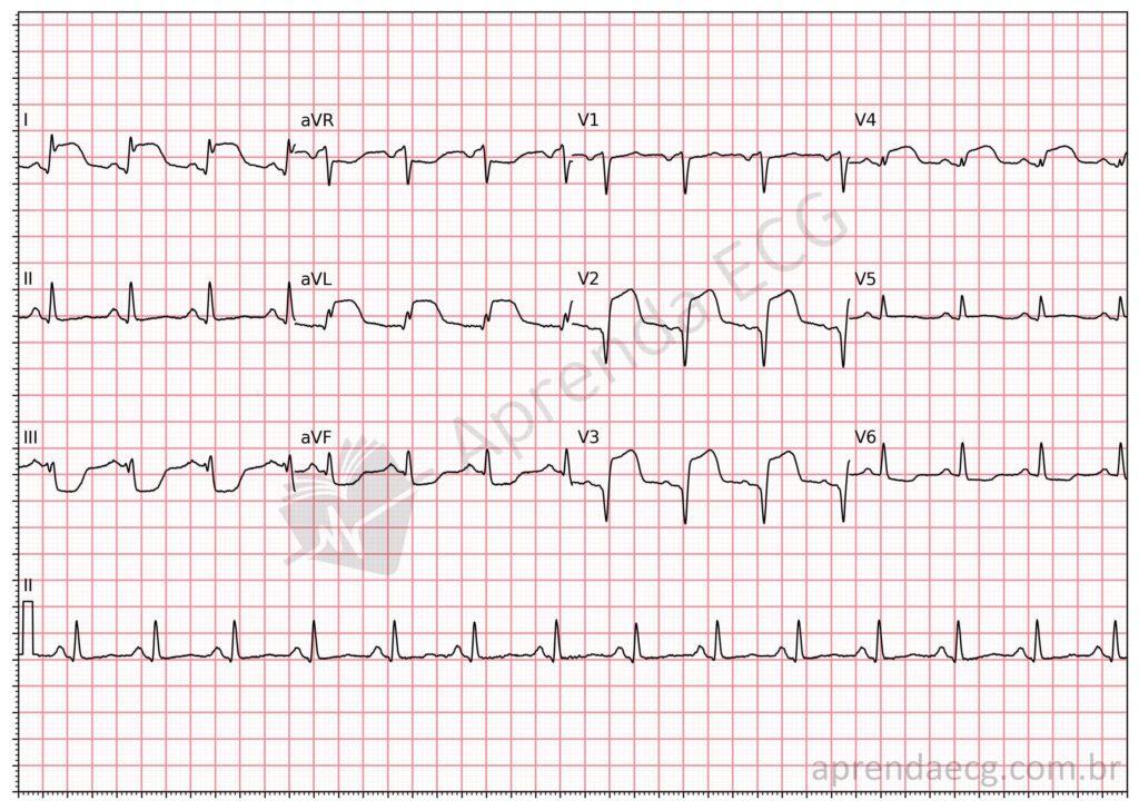
Historically, it was taught that concave morphology was “benign” and that convex morphology indicated ischemia. However, studies have shown that up to 43% of LAD occlusions present with concave morphology. Morphology, therefore, should not be used in isolation.
The evolution of morphology over time may be more informative: an ST segment that progresses from concave to convex reflects worsening transmural ischemia.
We have already covered this topic on our site in the article: The Myth of the Smiley-Face ST Elevation.
🔁 Reciprocal ST Depression: A Marker of Ischemia
Reciprocal, or “mirror-image”, ST depression is a strong indicator of transmural infarction and is present in up to 80% of STEMIs. Its presence is associated with a larger area of myocardium at risk and a worse prognosis. It also helps distinguish infarction from other conditions, such as pericarditis.
Reciprocal, or “mirror-image”, ST depression is an electrocardiographic change that reflects the remote repercussion of transmural ischemia. It arises from complex electrophysiological mechanisms involving vectors opposite to the site of myocardial injury, which makes it an indirect but valuable manifestation of acute ST-elevation infarction.
It is present in up to 80% of STEMIs and is associated with the extent of the ischemic area, a higher risk of ventricular dysfunction, and a worse clinical prognosis. In many cases, reciprocal ST depression is the first or only sign of significant ischemia in the territory opposite the affected lead.
Reciprocal ST depression increases the specificity of the diagnosis of MI and can help differentiate MI from pericarditis, since the latter usually shows diffuse ST elevation without reciprocal changes.
🗺️ Infarct Localization: Using the ECG as a Coronary Map
The topographic distribution of ST changes allows the culprit artery to be inferred. A systematic analysis of the leads makes it possible to identify septal, anterior, lateral, inferior, and RV infarctions. The use of additional leads (V7-V9, V3R-V4R) increases sensitivity for “hidden” infarctions.
| Terminology | Electrocardiographic Leads | Probable Site of Coronary Occlusion |
|---|---|---|
| Septal | V1 and V2 | Septal branch of the LAD |
| Anteroapical | V1-V2 through V3-V6 | Mid LAD |
| Extensive anterior | V1-V2 through V4-V6, I and aVL | Proximal LAD |
| Mid-anterior | I and aVL, sometimes V2-V3 | First diagonal branch |
| Lateral | I, aVL, V5 and V6 | Proximal LCx |
| Posterolateral | V7-V9, ST depression/RS in V1 and V2 | Proximal LCx |
| Inferior | II, III and aVF | RCA or LCx |
| Inferolateral | II, III, aVF, I, aVL, V5-V6, V7-V9 | RCA or LCx |
| Right ventricle | V3R and V4R | Proximal RCA |
⚠️ High-Risk Morphological Patterns
Some electrocardiographic patterns are associated with a worse prognosis:
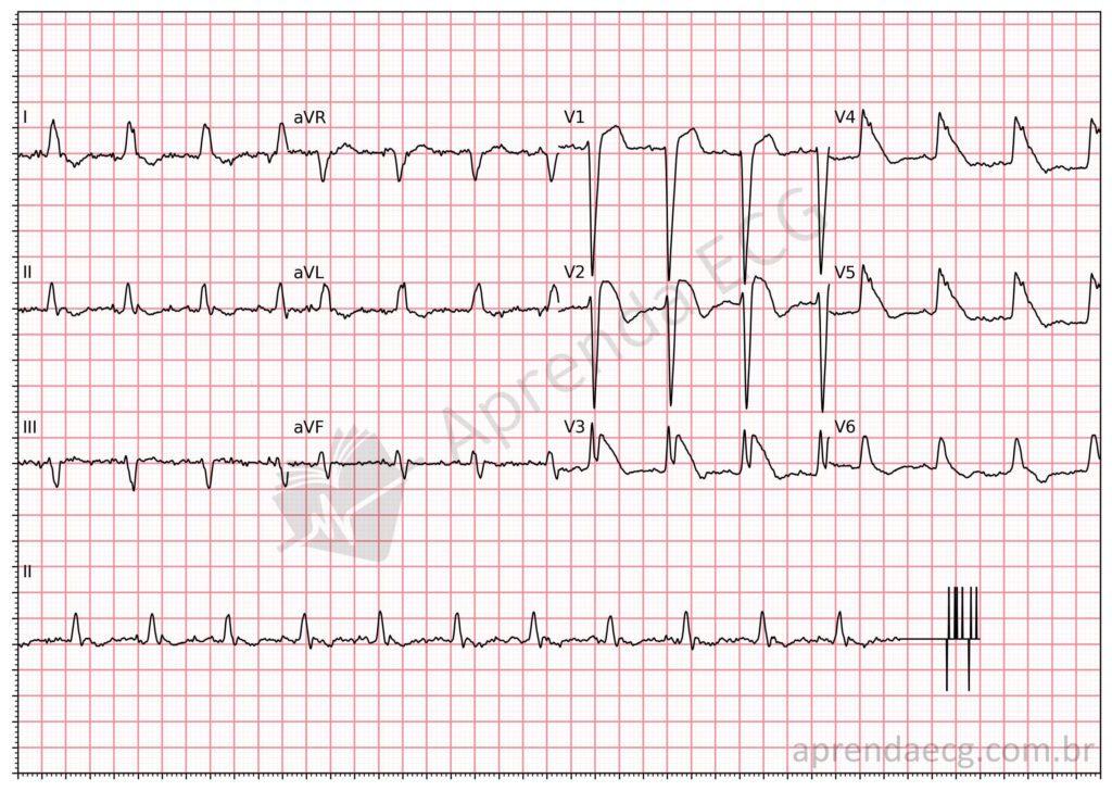
Tombstone Pattern
The tombstone pattern (“tombstoning ST elevation”) is a characteristic ST-elevation morphology associated with extensive acute infarctions, especially of the anterior wall, due to proximal occlusion of the left anterior descending artery (LAD).
Electrocardiographic features:
- Marked convex ST-segment elevation.
- The ST segment merges with the downstroke of the R wave or directly with a QS/QR complex.
- Absence of a clearly defined R wave, or an R wave < 40 ms in duration.
- The peak of the ST segment is higher than the amplitude of the R wave (when present).
- The T wave merges with the ST segment, forming a monophasic pattern.
Clinical implications:
- Associated with a large infarct area and an increased risk of cardiogenic shock and ventricular fibrillation.
- Increased mortality compared with other ST-elevation patterns.
- Requires an aggressive, immediate approach with early reperfusion.
Shark Fin Sign (lambda wave)
Also known as the “triangular pattern” or “giant R wave”, the shark fin sign is a rare but extremely serious ST-elevation morphology. The tracing resembles a fin emerging from the surface, the result of the fusion of the QRS complex, the ST segment, and the T wave.
Electrocardiographic features:
- Large-amplitude R wave (> 10 mm).
- Complete fusion of the QRS, ST, and T, forming a monophasic triangular morphology.
- Lambda wave: a prominent J component followed by a downsloping ST segment that merges with the negative T wave.
- Usually positive polarity in the leads facing the ischemic area.
Clinical implications:
- Strongly associated with OMI of the LAD or proximal RCA, with no collateral circulation.
- Indicator of high risk for ventricular fibrillation, out-of-hospital cardiac arrest, and cardiogenic shock.
- Reported in-hospital mortality between 50% and 70%.
📉 Sensitivity of ST Elevation
Despite being one of the most valued electrocardiographic criteria, ST-segment elevation has limited sensitivity for the diagnosis of acute coronary occlusion (OMI).
A recent meta-analysis performed by our team, including three large studies with more than 23,000 patients, showed that the sensitivity of ST elevation for detecting OMI is only 43.6%. This means that more than half of the patients with an occluded artery did not show classic ST elevation on the ECG.
On the other hand, specificity was quite high, reaching 96.5%, with a positive likelihood ratio (LR+) of 12.77, indicating that, when present, ST elevation substantially increases the probability of OMI. However, the negative likelihood ratio (LR–) was 0.58, showing that its absence is not enough to rule out occlusion MI.
These data highlight the importance of considering other electrocardiographic signs of occlusion, especially when the clinical picture suggests infarction but there is no classic ST elevation.
🔄 The New OMI/NOMI Paradigm
The traditional classification into STEMI and NSTEMI is based almost exclusively on the presence or absence of ST elevation. However, recent evidence shows that this approach has low sensitivity for detecting all acute coronary occlusions (OMI).
The OMI/NOMI paradigm views the ECG as a tool for detecting occlusion, regardless of the presence of “classic ST elevation”. This paradigm has already proven more sensitive, with an OMI detection rate of around 78% when multiple electrocardiographic signs are considered, while maintaining high specificity. In this way, patients with OMI but without classic ST elevation can be identified and treated early.
✅ Conclusion: ST Is Still King, But the Kingdom Is Bigger
ST-segment elevation remains a highly valuable marker, but it can no longer reign alone. Modern ECG interpretation requires recognition of morphological patterns, reciprocal signs, additional leads, and clinical correlation.
The absence of ST elevation does not rule out OMI. Recognizing “hidden” signs and high-risk patterns can make the difference between a well-managed patient and a missed diagnosis.
References
- Scheffer MK, De Marchi MFN, de Alencar Neto JN, Felicioni SP. Eletrocardiograma de A a Z. São Paulo: Manole, 2024.
- Pardee HEB. An electrocardiographic sign of coronary artery obstruction. Arch Intern Med. 1920;26(2):244–57.
- Byrne RA, Rossello X, Coughlan JJ, Barbato E, Berry C, Chieffo A, et al. 2023 ESC Guidelines for the management of acute coronary syndromes. Eur Heart J. 2023 Oct 12;44(38):3720–826.
- Thygesen K, Alpert JS, Jaffe AS, Chaitman BR, Bax JJ, Morrow DA, et al. Fourth universal definition of myocardial infarction (2018). Eur Heart J. 2018 Jan 14;40(3):237–69.
- Martí Sánchez D, Mestre JL, Salido L, Esteban MJ, Casas E, Pey J, et al. Incidence, angiographic features and outcomes of patients presenting with subtle ST-elevation myocardial infarction. Am Heart J. 2014;168(6):884–90.
- Martí D, Salido L, Mestre JL, Casas E, Esteban MJ, Zamorano JL. Usefulness of Reciprocal Changes in the Diagnosis of Myocardial Infarction With Minimal ST-segment Elevation. Rev Española Cardiol. 2016 Jul;69(7):706–7.
- Smith SW. Upwardly concave ST segment morphology is common in acute left anterior descending coronary occlusion. J Emerg Med. 2006 Jul;31(1):69–77.
- Vaidya GN, Antoine S, Imam SH, Kozman H, Smulyan H, Villarreal D. Reciprocal ST-Segment Changes in Myocardial Infarction: Ischemia at Distance Versus Mirror Reflection of ST-Elevation. Am J Med Sci. 2018;355(2):162–7.
- Wimalaratna HSK. “Tombstoning” of ST segment in acute myocardial infarction. Lancet. 1993 Aug;342(8869):496.
- Guo XH, Yap YG, Chen LJ, Huang J, Camm AJ. Correlation of coronary angiography with “tombstoning” electrocardiographic pattern in patients after acute myocardial infarction. Clin Cardiol. 2000;23(5):347–52.
- Cipriani A, D’Amico G, Brunello G, Perazzolo Marra M, Migliore F, Cacciavillani L, et al. The electrocardiographic “triangular QRS-ST-T waveform” pattern in patients with ST-segment elevation myocardial infarction: Incidence, pathophysiology and clinical implications. J Electrocardiol. 2018;51(1):8–14.
- de Alencar Neto JN, Scheffer MK, Correia BP, Franchini KG, Felicioni SP, De Marchi MFN. Systematic review and meta-analysis of diagnostic test accuracy of ST-segment elevation for acute coronary occlusion. Int J Cardiol. 2024 Feb;131889.