
ST-segment elevation is an electrocardiographic finding that can be seen in several clinical conditions, some of them unrelated to myocardial ischemia. The myth of the “smiley-face” ST elevation arose from the perception that ST elevation with a concave morphology indicated benign or non-ischemic conditions, while a convex morphology (the so-called “frowny-face” ST elevation) was associated with acute myocardial infarction (MI). However, this simplified distinction is not supported by the scientific evidence.
It is common for young people, especially men, to show ST-segment elevation in leads V2 and V3, which may range from 1 to 3 mm, without any pathological significance. This pattern, known as the male pattern, should not be confused with ischemia. However, a non-concave (convex or straight) ST-segment morphology is highly specific for ischemia and is seen in 97% of MI cases.
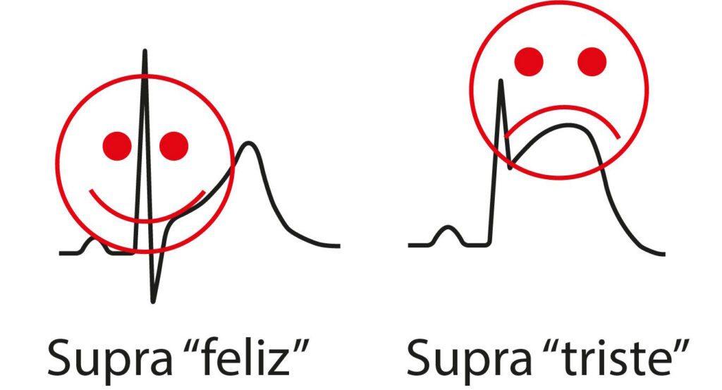
Debunking the Smiley-Face ST Elevation
The concept of the “smiley-face” ST elevation, which associates a concave ST-segment morphology with benign conditions, has been widely disseminated, but recent studies show that this distinction is not so clear. A study by Smith et al. revealed that concave morphology may be present in up to 43% of cases of acute occlusion of the left anterior descending artery (OMI). This finding shows that concave ST morphology should not automatically be dismissed as “benign”.
Another study by Kosuge et al. explored the relationship between the shape of ST-segment elevation and infarct size in patients with anterior MI. Although concave morphology was associated with smaller infarctions and better recovery of ventricular function, this does not rule out ischemia. Thus, ST morphology alone is not enough to determine the severity or benign nature of a cardiac condition.
The Importance of a Comprehensive Approach
Distinguishing ischemic from non-ischemic ST elevation remains a challenge in clinical practice. Although convex morphology is more specific for the diagnosis of ischemia, it is essential to consider other electrocardiographic signs, such as reciprocal ST depression, which is a robust marker of MI. In addition, the evolution of ST-segment morphology over time, from concave to convex, may reflect the progression of myocardial ischemia.
Therefore, the myth that a concave ST morphology (the “smiley face”) always indicates benign conditions is incorrect. ECG interpretation should be done together with a complete clinical assessment, taking into account other electrocardiographic findings and the temporal evolution of the changes.
The New OMI/NOMI Paradigm
Want to understand more about the complexities of diagnosing acute coronary occlusion and explore the new OMI/NOMI paradigm? We invite you to discover the latest approaches, which go beyond simple ST-segment morphology, and to stay up to date on advances in electrocardiogram interpretation. Visit our section on the new OMI/NOMI paradigm and sharpen your knowledge of cardiology!
References
- Hiss RG, Lamb LE, Allen MF. Electrocardiographic findings in 67,375 asymptomatic subjects. X. Normal values*. Am J Cardiol. 1960;6(1):200–31.
- Brady WJ, Syverud SA, Beagle C, Perron AD, et al. Electrocardiographic ST-segment elevation: The diagnosis of acute myocardial infarction by morphologic analysis of the ST segment. Acad Emerg Med. 2001;8(10):961–7.
- Smith SW. Upwardly concave ST segment morphology is common in acute left anterior descending coronary occlusion. J Emerg Med. 2006;31(1):69–77.
- Kosuge M, Kimura K, Ishikawa T, Kuji N, et al. Value of ST-segment elevation pattern in predicting infarct size and left ventricular function at discharge in patients with reperfused acute anterior myocardial infarction. Am Heart J. 1999;137(3):522–7.