

ST-segment elevation is an electrocardiographic pattern of transmural ischemia that appears within minutes of acute coronary occlusion. When it occurs in the setting of ACS, it is dynamic and usually limited to the territory of the culprit coronary artery. The presence of pathological Q waves or reciprocal ST depression supports the diagnosis of evolving infarction.
Electrocardiographic Criteria
- ST-segment elevation ≥ 1 mm in any lead except V2 and V3
- For V2 and V3: ≥ 2 mm in men aged 40 years or older; ≥ 2.5 mm in men younger than 40 years; ≥ 1.5 mm in women of any age
Applicable in the absence of LVH or LBBB.
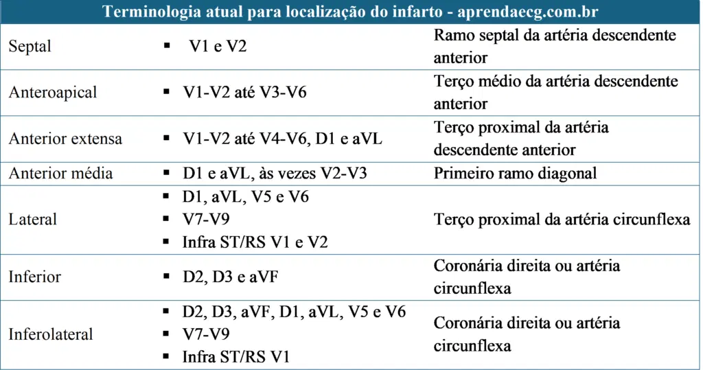
Infarct Topography
Concave vs. Convex ST Elevation
Convex ST-segment morphology is highly specific for the diagnosis of MI; however, it can evolve from concave to straight and finally to convex. It has been reported that 43% of patients undergoing primary angioplasty of the left anterior descending artery had a concave pattern in the anterior leads, leading to the conclusion that this feature cannot be used to rule out ACS.
Reciprocal ST-Segment Depression
ST-segment depression may be seen in leads opposite to those showing ST elevation and is called reciprocal or mirror-image change.
ECG in ST-Segment Elevation of Ischemic Origin
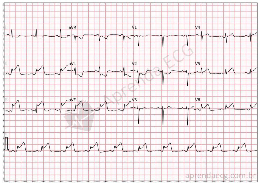
ST-segment elevation: acute inferolateral wall infarction. Note the ST-segment elevation in the inferolateral wall (II, III, aVF, V5 and V6) with reciprocal ST depression in the mid-anterior wall (I, aVL and V2). Pattern consistent with acute coronary occlusion.
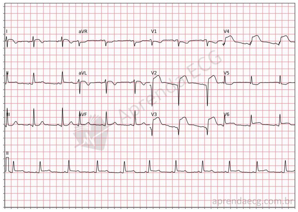
ST-segment elevation: acute extensive anterior wall infarction. Note the ST-segment elevation in the extensive anterior wall (V2-V6, I and aVL). Pathological Q waves are also present from V2 to V4. Pattern consistent with acute coronary occlusion.
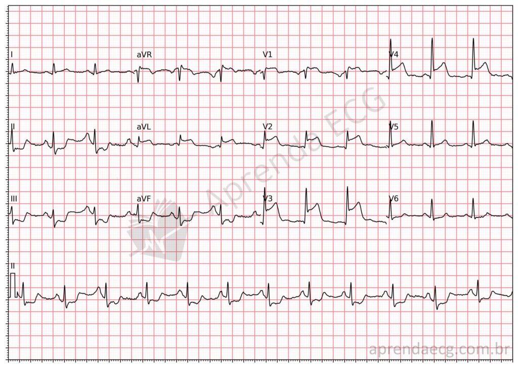
ST-segment elevation: acute extensive anterior wall infarction. Note the ST-segment elevation in the extensive anterior wall (V1-V4 and aVL) with reciprocal ST depression in the inferior wall (II, III and aVF). Pattern consistent with acute coronary occlusion.
Other Electrocardiographic Signs of OMI
Remember that, given the low sensitivity of ST-segment elevation for the diagnosis of acute coronary occlusion (OMI) (43.6%), ECG analysis should also consider other signs that indicate occlusion.
References
- Scheffer MK, De Marchi MFN, de Alencar Neto JN, Felicioni SP. Eletrocardiograma de A a Z. São Paulo: Manole, 2024.
- Alencar JN De, Feres F, Marchi MFN De, Franchini KG, Scheffer MK, Felicioni SP, et al. Além do Paradigma IAMCSST-IAMSSST: Proposta do Instituto Dante Pazzanese para o Diagnóstico de Oclusão Coronariana Aguda. Arq Bras Cardiol. 2024;121(5):e20230733.
- de Alencar Neto JN, Scheffer MK, Correia BP, Franchini KG, et al. Systematic review and meta-analysis of diagnostic test accuracy of ST-segment elevation for acute coronary occlusion. Int J Cardiol. 2024;131889.
- Aslanger EK, Yıldırımtürk Ö, Şimşek B, Bozbeyoğlu E, et al. DIagnostic accuracy oF electrocardiogram for acute coronary OCClUsion resuLTing in myocardial infarction (DIFOCCULT Study). IJC Hear Vasc. 2020;30.
- Thygesen K, Alpert JS, Jaffe AS, Chaitman BR, et al. Fourth universal definition of myocardial infarction (2018). Eur Heart J. 2018;40(3):237–69.
- Huang HD, Birnbaum Y. ST elevation: Differentiation between ST elevation myocardial infarction and nonischemic ST elevation. J Electrocardiol. 2011;44(5):494.e1-494.e12.
- Nable JV, Brady W. The evolution of electrocardiographic changes in ST-segment elevation myocardial infarction. Am J Emerg Med. 2009;27(6):734–46.
- Smith SW. Upwardly concave ST segment morphology is common in acute left anterior descending coronary occlusion. J Emerg Med. 2006;31(1):69–77.
- Bayés de Luna A. New heart wall terminology and new electrocardiographic classification of Q-wave myocardial infarction based on correlations with magnetic resonance imaging. Rev Esp Cardiol. 2007;60(7):683–9.
- Bayés de Luna A. Location of Q-wave myocardial infarction in the era of cardiac magnetic resonance imaging techniques: An update. J Electrocardiol. 2007;40(1):69–71.


