

Dear reader, do you remember that ECG lecture with blue-background slides, yellow text and a big title: “Ischemia, Injury and Necrosis”? Among the patterns described, one of them was (with emphasis on the past tense) Subepicardial Ischemia. This pattern was defined by negative, symmetric T waves in two contiguous leads, and it still appears in guidelines today.
Why “Subepicardial Ischemia” Doesn't Add Up (Not Even Pathophysiologically)
Originally, inverted primary T waves (symmetric or nearly symmetric) were attributed to a specific type of ischemia that supposedly affected only the subepicardial region. Pathophysiologically, however, the first cardiac layer to become ischemic when coronary blood flow is interrupted is the one farthest from the coronary arteries (the endocardium), not the closest (the epicardium).
Supporting this pathophysiological view with cardiac magnetic resonance findings, a consensus written by leading figures in electrocardiography such as Bayés de Luna, Hein Wellens et al. noted that, in general, isolated T-wave inversion does not occur during ongoing acute ischemia and, in particular, isolated subepicardial ischemia does not occur. In fact, what causes T-wave inversion in the ischemic setting is transmural myocyte edema after reperfusion, not restricted to the subepicardium.
If you'd like an editorial “wake-up call” about this kind of label that has survived since the PowerPoint era, read this Aprenda ECG piece: ECG Myths: We Learned to Memorize Tracings, Not to Understand Them.
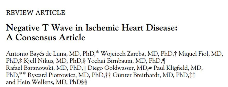
T-Wave Inversion: Primary vs. Secondary
An inverted T wave may be permanent or transient and may have a primary or secondary morphology. A secondary inverted T wave is associated with abnormal depolarization, as occurs for example in preexcitation, bundle branch block or the left ventricular strain pattern.
Primary T-wave inversion, by contrast, is caused by a change in repolarization itself, which may or may not be ischemic in origin. When it is ischemic, however, isolated T-wave inversion suggests that the evolving ischemia has been interrupted by reperfusion/recanalization, especially in the absence of pain.
How Primary Ischemic T-Wave Inversion Usually Appears
- The inverted T wave may be transient or persist for several days after the ischemic event or revascularization.
- An inverted T wave in one wall may be the reflection of a symmetric T wave with a large area relative to the QRS (hyperacute) in the opposite wall.
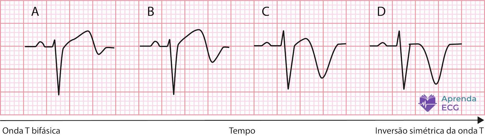
- The T wave may be terminally rather than completely inverted, with a positive initial portion (plus-minus morphology).
- A deeply inverted T wave may be accompanied by a U wave, which in ischemia usually has the same polarity as the T wave.
Clinical Context Rules the Report (With Pain vs. Without Pain)
Correct ECG interpretation always depends on the clinical context, and T-wave inversion is no exception. It is therefore essential to confirm whether the patient has cardiac pain at the time of the ECG, since its presence or absence should lead us to different conclusions.
As described in the Bayés de Luna consensus, a primary inverted T wave by itself does not indicate acute ischemia but rather subacute ischemia (as in reperfusion/recanalization). Thus, an inverted, symmetric T wave would represent acute ischemia only when it is the mirror image of a hyperacute T wave, playing a role analogous to that of ST depression as the mirror image of ST elevation.
Therefore, in acute ischemia, the inverted T wave is a supporting actor, not the lead.
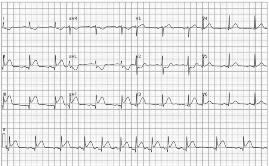
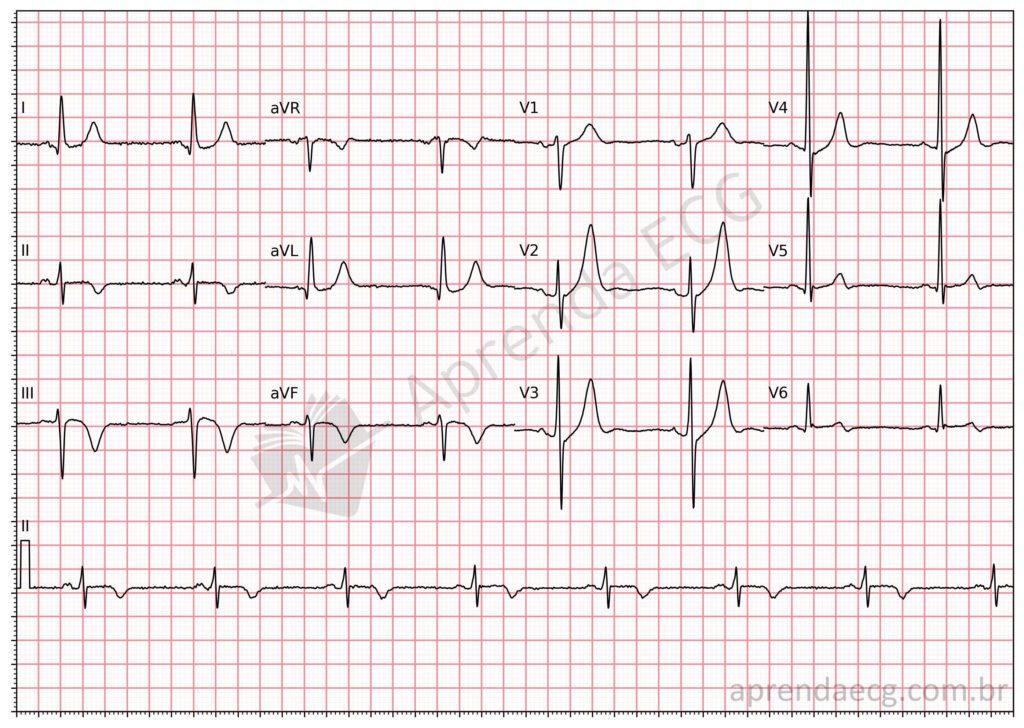
On an ECG recorded in a subacute anginal presentation, that is, in a patient who no longer has cardiac pain at the time of the test, the T wave may appear inverted or terminally inverted, representing recanalization/reperfusion (reperfusion T waves). Such reperfusion occurs after the previously occluded coronary artery opens, whether after vasospasm, as a result of an intervention (thrombolysis or primary angioplasty) or even spontaneously.
Wellens Syndrome: ECG Pattern and Classic Pitfalls
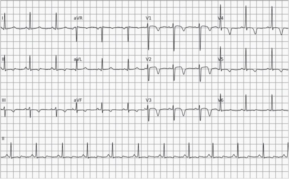
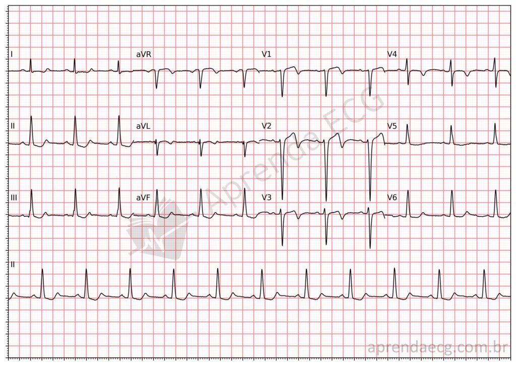
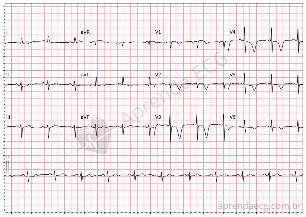
This occurs, for example, in Wellens syndrome , whose electrocardiographic pattern involves primary inverted T waves in the anterior leads. It should be stressed, however, that reperfusion T waves are not restricted to the anterior wall and can be seen in other coronary territories (for example, the inferior wall).
“Pattern A” vs. “Pattern B”: Why This Dichotomy Gets in the Way
Although the morphologies differ, there is no pathophysiological difference between terminal and complete T-wave inversion in the subacute ischemic setting: one is simply the temporal evolution of the other. The distinction between Wellens “pattern A” and “pattern B” should therefore be avoided.
References
- Scheffer MK, de Alencar Neto JN, De Marchi MFN, Felicioni SP. Eletrocardiograma na síndrome coronária aguda. São Paulo: Manole; 2025.
- Scheffer MK, De Marchi MFN, de Alencar Neto JN, Felicioni SP. Eletrocardiograma de A a Z. São Paulo: Manole; 2024.
- Driver BE, Shroff GR, Smith SW. Posterior reperfusion T-waves: Wellens’ syndrome of the posterior wall. Emerg Med J. 2017;34(2):119-123. doi:10.1136/emermed-2016-205852.
- de Luna AB, Zareba W, Fiol M, Nikus K, Birnbaum Y, Baranowski R, et al. Negative T wave in ischemic heart disease: a consensus article. Ann Noninvasive Electrocardiol. 2014;19(5):426-441. doi:10.1111/anec.12193.