

The Wellens electrocardiographic pattern is associated with impending MI, mainly with a proximal left anterior descending artery lesion as the culprit.
Electrocardiographic Criteria
- Isoelectric or minimally elevated ST segment (up to 1 mm) with a biphasic T wave (“plus-minus”) or a deep, symmetrically inverted T wave in leads V2 and V3
ECG in Wellens Syndrome
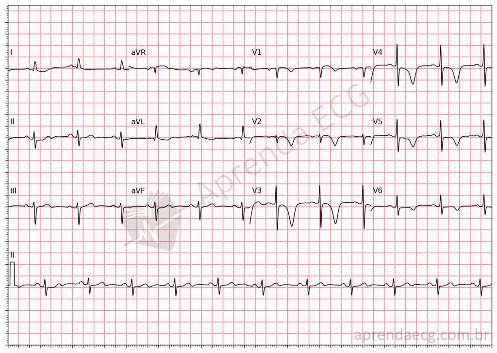
Wellens pattern. On this ECG we see the Wellens pattern with negative, symmetric T waves in the precordial leads.
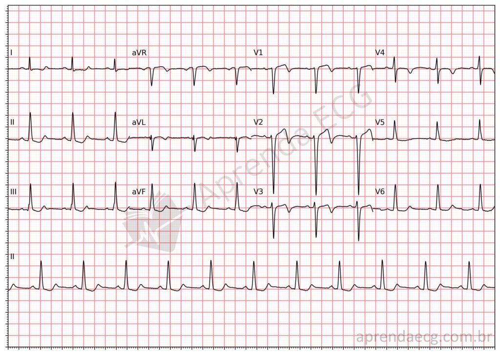
Wellens pattern. On this tracing, in contrast, we see the plus-minus T-wave pattern, that is, with an initial positive portion and a terminal negative portion in precordial leads V2 and V3.
Related Topics
References
- de Zwaan C, Bär FWHM, Wellens HJJ. Characteristic electrocardiographic pattern indicating a critical stenosis high in left anterior descending coronary artery in patients admitted because of impending myocardial infarction. Am Heart J. 1982;103(4 PART 2):730–6.
- Kobayashi A, Misumida N, Aoi S, Kanei Y. Prevalence and Clinical Implication of Wellens’ Sign in Patients With Non-ST-Segment Elevation Myocardial Infarction. Cardiol Res. 2019;10(3):135–41.
- Zhou L, Gong X, Dong T, Cui H he, et al. Wellens’ syndrome: incidence, characteristics, and long-term clinical outcomes. BMC Cardiovasc Disord. 2022;22(1):1–8.


