
Conduction disturbances in acute myocardial infarction (MI) are more than transient electrical complications. They are often anatomical markers of the involved territory, of the extent of the area at risk and, in certain settings, of prognosis. Correct interpretation of these disturbances requires integrating coronary anatomy, the blood supply of the conduction system, and clinical electrophysiology.
In the era of primary angioplasty, the incidence of advanced blocks has decreased, but their prognostic significance remains relevant. Understanding where the block occurs, suprahisian or infrahisian, is essential to anticipate the clinical course.
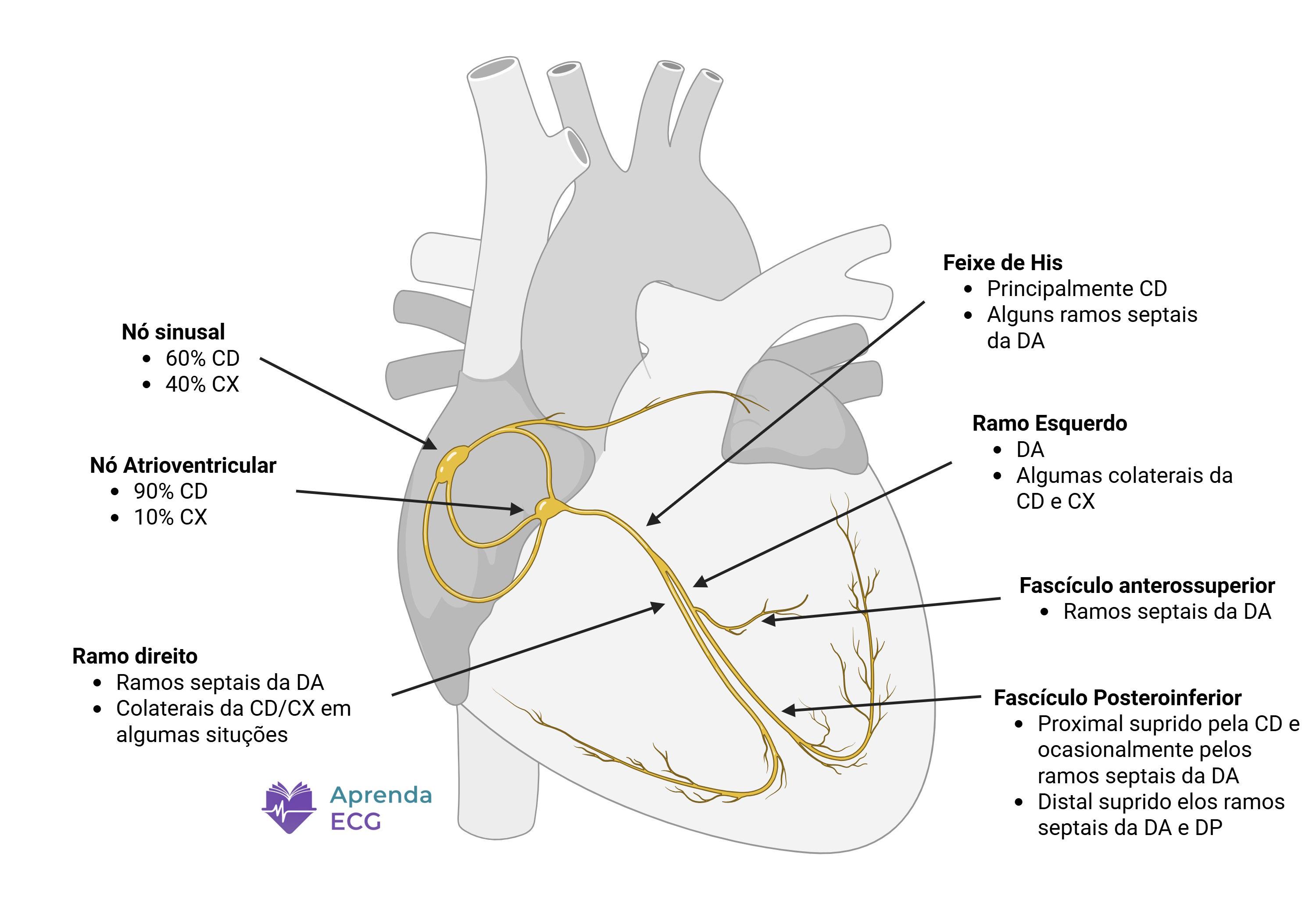
Blood Supply of the Conduction System
The vascular distribution of the conduction system explains most of the patterns seen in MI.
- Sinoatrial node (SAN): supplied by the right coronary artery (RCA) in about two thirds of individuals and by the circumflex artery (LCx) in the rest.
- Atrioventricular node (AVN): supplied, in most cases, by the RCA through the AV nodal branch; in left-dominant circulation, by the LCx.
- His bundle and proximal bundle branches: dual supply, with contributions from the AV nodal branch (proximal portion) and from the septal branches of the left anterior descending artery (LAD).
- Right bundle branch and left anterior fascicle: depend predominantly on the septal branches of the LAD.
- Left posterior fascicle: more robust and often dual supply (LAD and RCA), which explains its lower vulnerability.
This anatomical organization establishes a direct relationship between the infarcted territory and the type of block observed.
Inferior MI and Suprahisian Disturbances
Inferior MI is classically associated with bradyarrhythmias and atrioventricular blocks (AV blocks) at the suprahisian level.
Two main mechanisms are involved:
- AVN ischemia, due to RCA occlusion proximal to the nodal branch.
- Vagal hypertonia (Bezold-Jarisch reflex), especially in the first hours of the event.
Typical manifestations
- Sinus bradycardia
- First-degree AV block
- Second-degree Mobitz type I AV block (Wenckebach)
- Complete AV block with narrow QRS and junctional escape rhythm
The electrocardiographic pattern usually progresses gradually (prolonged PR → Wenckebach → complete AV block), with narrow QRS complexes, suggesting a nodal block.
In the pre-reperfusion era, advanced AV blocks in inferior MI could occur in up to 15–20% of cases. Contemporary studies show a significantly lower incidence after primary PCI, generally below 3%.
Prognostic implications
In inferior MI, advanced AV block is usually transient and resolves with reperfusion. The need for temporary pacing is restricted to cases with hemodynamic instability or an inadequate escape rhythm.
When the block is nodal with a narrow QRS, the prognosis is generally favorable and reflects vagal activation or transient AVN ischemia rather than extensive necrosis of the His-Purkinje system.
Anterior MI and Infrahisian Disturbances
The scenario changes completely in anterior MI, especially when there is proximal LAD occlusion before the origin of the first septal branch.
In this setting, the His-Purkinje system is directly compromised.
Typical manifestations
- New right bundle branch block (RBBB)
- Left anterior fascicular block (LAFB)
- Bifascicular block (RBBB + LAFB)
- Advanced AV block with wide QRS and ventricular escape rhythm
Unlike inferior MI, AV block in anterior MI usually appears suddenly, without gradual PR prolongation, and with a widened QRS, suggesting an infrahisian block.
This pattern reflects extensive necrosis of the interventricular septum and is associated with greater left ventricular dysfunction.
Anatomical significance
New RBBB in the setting of chest pain is highly suggestive of proximal LAD occlusion, since the right bundle branch depends directly on the septal branches.
When combined with LAFB, the pattern indicates extensive septal involvement. In practice, this combination works as an electrical marker of proximal occlusion.
Prognosis
Unlike the nodal block of inferior MI, infrahisian block in anterior MI is associated with:
- Larger area of necrosis
- Higher incidence of cardiogenic shock
- Higher in-hospital mortality
- Higher likelihood of requiring a permanent pacemaker
Here, the block is not merely an electrical epiphenomenon: it is a marker of extensive infarction.
Left Bundle Branch Block in the Setting of MI
A left bundle branch block (LBBB) was historically considered a STEMI equivalent when new or presumably new.
However, contemporary evidence shows that LBBB chronology alone has low diagnostic value. Distinguishing new from old LBBB has a low likelihood ratio and does not significantly change the post-test probability of coronary occlusion.
On the other hand, specific electrocardiographic criteria for ischemia in LBBB, such as the modified Sgarbossa criteria, have substantially better diagnostic performance and should guide clinical decisions.
From an anatomical standpoint, new LBBB in MI suggests multivessel disease or extensive septal involvement, since the left bundle branch has a relatively protected and extensive blood supply.
Incidence and Impact in the Primary PCI Era
With the widespread adoption of primary angioplasty, the incidence of advanced AV block has decreased significantly compared with the 1960s–1980s.
Even so, when present, conduction disturbances remain associated with a worse prognosis, mainly in the following scenarios:
- Anterior MI with infrahisian block
- LBBB associated with ischemic signs
- Bifascicular block in extensive infarction
Importantly, in inferior MI, the presence of advanced AV block does not always translate into higher mortality after multivariable adjustment, unlike anterior MI.
Thus, the prognostic significance depends more on the territory than on the block itself.
Clinical Integration: What Does the Block Tell Us?
In practical terms:
Conclusion
Conduction disturbances in MI should be interpreted as anatomical and prognostic markers, not merely as electrical complications.
Inferior MI tends to produce suprahisian blocks, usually transient and with a better prognosis. Anterior MI, especially from proximal LAD occlusion, compromises the His-Purkinje system and is associated with infrahisian blocks, greater extent of necrosis, and a worse clinical course.
References
- Nikus K, Birnbaum Y, Fiol-Sala M, Rankinen J, Bayés de Luna A. Conduction Disorders in the Setting of Acute STEMI. Curr Cardiol Rev. 2021;17:41-49.
- Ibanez B, James S, Agewall S, et al. 2017 ESC Guidelines for the management of acute myocardial infarction in patients presenting with ST-segment elevation. Eur Heart J. 2018;39(2):119-177. doi:10.1093/eurheartj/ehx393.
- Kosmidou I, Redfors B, Dordi R, et al. Incidence, Predictors, and Outcomes of High-Grade Atrioventricular Block in Patients With ST-Segment Elevation Myocardial Infarction Undergoing Primary Percutaneous Coronary Intervention (from the HORIZONS-AMI Trial). Am J Cardiol. 2017;119(9):1295-1301. doi:10.1016/j.amjcard.2017.01.019.
- Kim KH, Jeong MH, Ahn Y, et al. Differential Clinical Implications of High-Degree Atrioventricular Block Complicating ST-Segment Elevation Myocardial Infarction according to the Location of Infarction in the Era of Primary Percutaneous Coronary Intervention. Korean Circ J. 2016;46(3):315-323. doi:10.4070/kcj.2016.46.3.315.
- de Alencar JN, Lima GWF, Geraldo HAS, et al. Accuracy of Left Bundle Branch Block Chronology and Electrocardiography Criteria for Acute Myocardial Infarction Diagnosis: A Systematic Review and Meta-analysis. Arq Bras Cardiol. 2025;122(10):e20250109. doi:10.36660/abc.20250109.
- Bayés de Luna A, Fiol-Sala M. Conduction disturbances in acute myocardial infarction. In: The ECG in ischemic heart disease. Blackwell Futura; 2008.