The Wenckebach phenomenon is one of the types of atrioventricular block that most intrigues cardiologists. Identifying it is essential to understand the electrical behavior of the heart in various clinical situations. In this post, we will explore in detail how the Wenckebach phenomenon appears on the electrocardiogram (ECG) and what its clinical implications are. Read to the end to understand how to differentiate it from other types of block and why this phenomenon matters in medical practice.
What Is the Wenckebach Phenomenon?
The Wenckebach phenomenon, also known as second-degree atrioventricular block type I, or Mobitz I, is characterized by progressive slowing of conduction between the atria and the ventricles, eventually leading to failure to conduct an atrial impulse. This failure results in a ventricular pause that is shorter than twice the preceding R-R interval, which contributes to the cyclical behavior typical of this phenomenon.
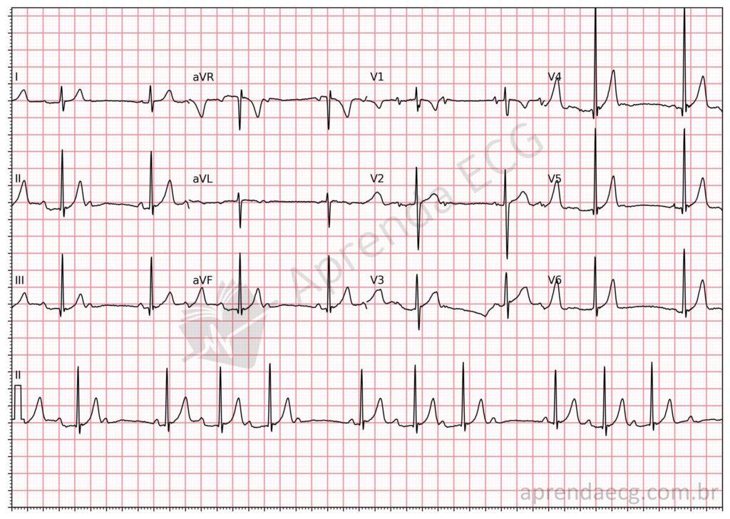
On the ECG, Wenckebach manifests as progressive prolongation of the PR interval until a QRS complex is dropped, after which the cycle starts again. This phenomenon occurs because of increasing conduction time in the atrioventricular (AV) node, where each successive impulse finds the AV node in a more refractory phase, resulting in progressively slower conduction until an impulse is not conducted.
Features of the Wenckebach Phenomenon
To better understand the Wenckebach phenomenon, it is important to recognize its main ECG features:
- Progressive PR Prolongation: with each beat, the PR interval gradually lengthens until a beat is dropped.
- Pause Shorter Than Twice the R-R Interval: the pause between QRS complexes after the block is shorter than twice the preceding R-R interval.
- Progressive Shortening of the R-R Interval: during the Wenckebach cycle, the R-R intervals shorten until the block occurs.
- Group Beating: the Wenckebach phenomenon is often described as showing “group beating” or “grouped conduction”, with repeated cycles of progressively longer PR intervals and an eventual conduction failure.
These features make the Wenckebach phenomenon an easy pattern for experienced cardiologists to identify, but it can be confused with other types of block, such as Mobitz II, especially in atypical cases. It is important to remember that in Wenckebach the block occurs predominantly in the AV node, whereas Mobitz II is usually associated with the His-Purkinje system.
Clinical Implications of the Wenckebach Phenomenon
The Wenckebach phenomenon is usually benign and is frequently found in young individuals and athletes, especially during sleep or in situations of increased vagal tone. Under these conditions, the heart's adaptation to increased parasympathetic tone results in a functional, physiological block that rarely requires intervention.
However, when seen in older patients or associated with symptoms such as dizziness, syncope, or heart failure, the Wenckebach phenomenon may indicate significant AV node disease or underlying structural heart disease. In these situations, further evaluation is needed, and a pacemaker may be considered to prevent prolonged pauses and syncope.
Atypical Wenckebach: A Diagnostic Challenge
Not all cases of Wenckebach follow the typical pattern described above. Atypical cases are particularly common in patients with dual AV nodal physiology or under the effect of high vagal stimulation. In such cases, the pattern of PR prolongation may be irregular, making the diagnosis more challenging. This highlights the importance of careful analysis of the PR-RR cycles and, when necessary, an electrophysiological study to confirm the diagnosis.
Key Points
- The Wenckebach phenomenon is a type of second-degree atrioventricular block characterized by progressive prolongation of the PR interval followed by a conduction failure.
- It is usually benign and commonly found in young individuals and athletes, but it may have significant clinical implications in older or symptomatic patients.
- Differential diagnosis with other types of block, such as Mobitz II, is essential for the correct therapeutic approach.
- In atypical cases, ECG interpretation can be challenging and requires a detailed understanding of the electrophysiological behavior of the AV node.
References
- Scheffer MK, De Marchi MFN, de Alencar Neto JN, Felicioni SP. Eletrocardiograma de A a Z. São Paulo: Manole, 2024.
- Issa Z, Miller J, Zipes D. Clinical Arrhythmology and Electrophysiology: A Companion to Braunwald’s Heart Disease. 3a ed. Philadelphia: Elsevier; 2019. 752 p.
- Mond HG, Vohra J. The Electrocardiographic Footprints of Wenckebach Block. Heart, Lung and Circulation. 2017.
- Hansom SP, Golian M, Green MS. The Wenckebach Phenomenon. Current Cardiology Reviews. 2021.