Atrioventricular (AV) block is traditionally classified as first, second, and third degree. This classification is useful from a descriptive standpoint. However, it does not answer the most important physiological question: where is the block?
The functional anatomy of the conduction system determines the electrocardiographic behavior of AV blocks. Mobitz I, Mobitz II, 2:1, or complete block are electrical expressions of distinct properties of the AV node and the His-Purkinje system. Understanding these properties allows the anatomical location of the disturbance to be inferred more precisely.
This article has an exclusively conceptual focus: to discuss how the electrophysiological characteristics of each segment of the conduction system explain the different block patterns seen on the ECG.
Functional anatomy of the conduction system
The electrical impulse travels through an organized sequence:
- Sinus node
- Atrial myocardium
- Atrioventricular node (AV node)
- His bundle
- Right and left bundle branches
- Purkinje network
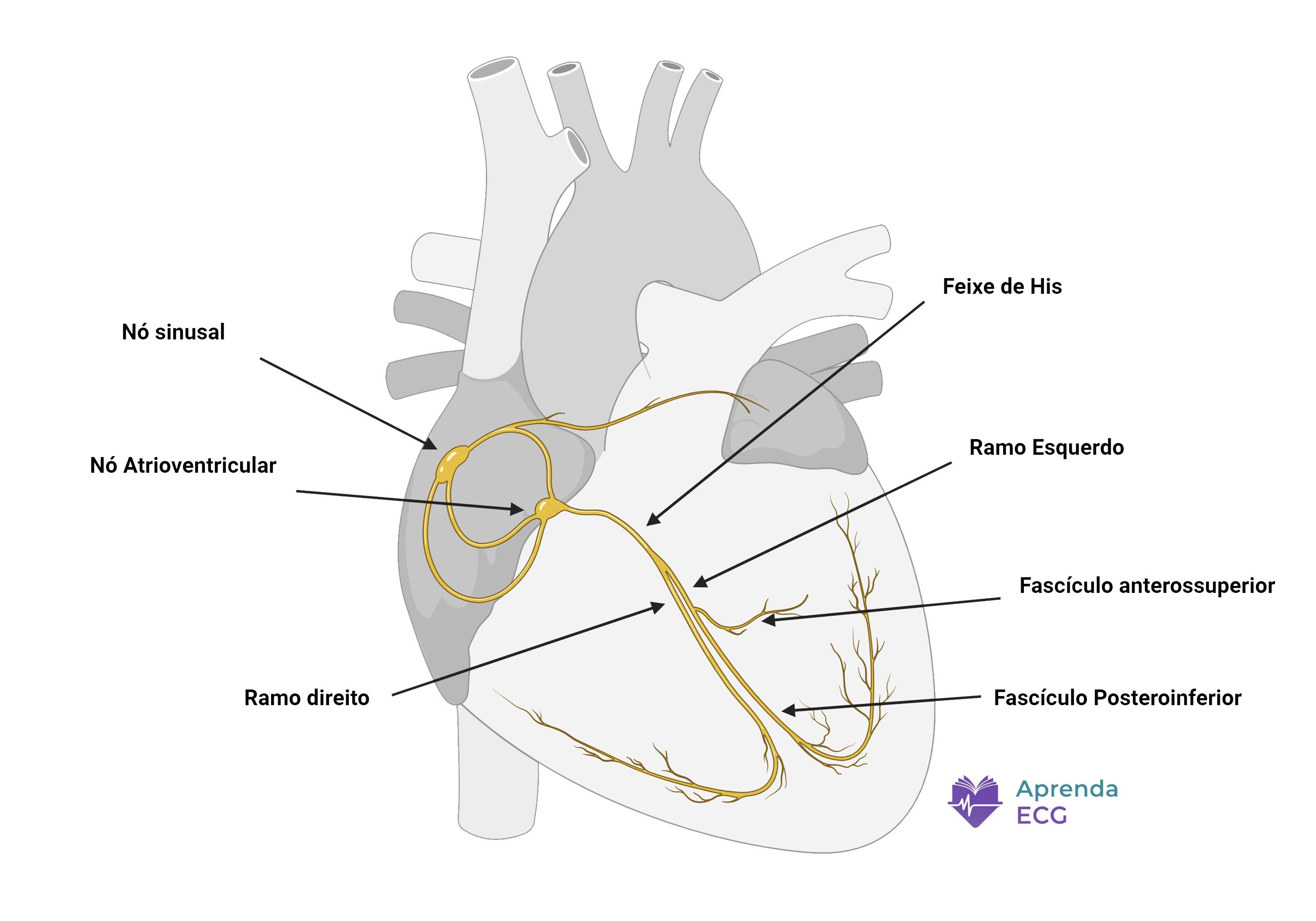
From an anatomical and electrophysiological standpoint, when thinking about AV block the system can be divided into three major territories:
- Suprahisian (nodal)
- Intrahisian
- Infrahisian (distal His-Purkinje system)
Each of these segments has its own conduction properties.
The AV node: decremental conduction
The AV node is composed of fibers with slow, calcium-dependent conduction. Its main electrophysiological feature is decremental conduction: the earlier or more frequent the impulse, the greater the conduction delay.
- Filter high atrial rates
- Ensure adequate ventricular filling time
- Allow rapid autonomic adaptation
On the surface ECG, AV node activity is represented mostly by the PR interval.
The His-Purkinje system: fast, non-decremental conduction
Unlike the AV node, the His-Purkinje system shows:
- Fast conduction
- Dependence on fast sodium channels
- “All-or-none” behavior
Under physiological conditions, there is no significant decremental conduction in this territory. When block occurs, it tends to be sudden, without prior progressive prolongation.
Nodal (suprahisian) block
Nodal block occurs within the AV node or at the proximal node-His junction.
Physiological basis
Because the AV node shows decremental conduction, a progressive increase in conduction delay is a natural consequence of its physiology. Thus, when we see:
- Progressive PR prolongation
- Grouped beats
- Pauses shorter than two preceding RR intervals
we are seeing the electrical expression of a nodal property.
The Wenckebach phenomenon is therefore the classic manifestation of nodal block.
Typical electrocardiographic expression
- Narrow QRS
- Variable or progressively prolonged PR interval
- Junctional escape rhythm in cases of complete nodal block
Conceptually, nodal block reflects a quantitative change in conduction (slower), not an abrupt structural failure.
Intrahisian block: the intermediate zone
Between the AV node and the ventricular bundle branches lies a short but functionally relevant segment: the His bundle.
Intrahisian block is less intuitive on the surface ECG. It may show mixed features:
- Fixed or slightly prolonged PR
- Narrow or slightly widened QRS
- Patterns that mimic Mobitz I or Mobitz II
The conventional ECG does not directly record His bundle activity. A definitive distinction between nodal and intrahisian block often requires an invasive electrophysiology study.
Infrahisian block (His-Purkinje system)
Infrahisian block involves the right and left bundle branches or the distal Purkinje system.
Physiological basis
Because the His-Purkinje system does not show significant decremental conduction, its failure tends to be abrupt. Thus, on the ECG we see:
- Fixed PR interval before the failure
- A sudden non-conducted P wave
- No prior progressive prolongation
This behavior is typical of the pattern described as Mobitz II.
Complete atrioventricular block and the origin of the escape
In third-degree AV block, the anatomical location can be inferred from the morphology and rate of the escape rhythm.
- Junctional escape: narrow QRS, higher rate → suggests nodal block
- Ventricular escape: wide QRS, lower rate → suggests infrahisian block
The more distal the block, the slower the escape rhythm tends to be.
The 2:1 pattern and the limits of the ECG
In 2:1 block, one of every two P waves is conducted. It is not possible to see whether there would be progressive PR prolongation, which prevents direct classification as Mobitz I or II.
Anatomically, 2:1 block can be nodal, intrahisian, or infrahisian.
Anatomical inference: probabilistic reasoning
- Decremental conduction suggests the AV node
- Sudden failure with a fixed PR suggests the His-Purkinje system
- A wide QRS increases the probability of distal block
- A slow ventricular escape rhythm suggests infrahisian block
Anatomical localization of AV block on the ECG is probabilistic, based on the electrophysiological properties of each segment.
Conclusion
The traditional classification describes what we see on the ECG. Anatomical localization explains why we see it.
Asking “where is the block?” means replacing electrocardiographic description with physiological reasoning.
References
- Scheffer MK, De Marchi MFN, de Alencar Neto JN, Felicioni SP. Eletrocardiograma de A a Z . São Paulo: Manole; 2024.
- Samesima N, God EG, Kruse JCL, Leal MG, et al. Diretriz da Sociedade Brasileira de Cardiologia sobre a Análise e Emissão de Laudos Eletrocardiográficos – 2022 . Arq Bras Cardiol. 2022;93(3 Suppl 2):2–19.
- Kusumoto FM, Schoenfeld MH, Barrett C, Edgerton JR, et al. 2018 ACC/AHA/HRS Guideline on the Evaluation and Management of Patients With Bradycardia and Cardiac Conduction Delay . J Am Coll Cardiol. 2019;74(7):e51–156.
- Issa Z, Miller J, Zipes D. Clinical Arrhythmology and Electrophysiology: A Companion to Braunwald’s Heart Disease . 3rd ed. Philadelphia: Elsevier; 2019.