

Pulmonary embolism (PE) can cause a range of changes on the electrocardiogram (ECG), although the test has low sensitivity and specificity for diagnosing this condition. The most common changes reflect acute right ventricular strain, hypoxia, and changes in the heart's electrical conduction.
Electrocardiographic Changes
- Sinus tachycardia
- S1Q3T3 pattern
- RBBB
- Right QRS axis deviation
- ST-segment elevation
- Negative T waves in the anterior leads
Electrocardiographic changes have an obvious diagnostic limitation for PE: the ECG is not the test that diagnoses the disease. The tests that diagnose it are invasive or noninvasive contrast imaging studies of the pulmonary arteries.
ECG in Pulmonary Embolism
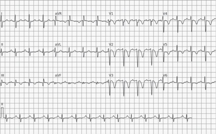
Pulmonary embolism. Example of an ECG with the S1Q3T3 pattern in a patient with a clinical picture suggestive of PE. The diagnosis was later confirmed by chest CT angiography.
Further Reading
Related Topics
References
- Alencar JN. Tratado de ECG. 2022.
- McGinn S, White PD. Acute cor pulmonale resulting from pulmonary embolism. J Am Med Assoc. 1935;104(17):1473.


