Pulmonary embolism (PE) is a potentially fatal medical emergency with a significant mortality rate. Although the definitive diagnosis depends on CT pulmonary angiography, the ECG can provide clues by revealing changes frequently associated with this condition. Among these changes, the famous S1Q3T3 pattern stands out, and it has particularities worth exploring.
Electrocardiographic Criteria
- S wave in lead I
- Q wave in lead III
- Inverted T wave in lead III
ECG with the S1Q3T3 Pattern
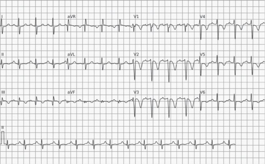
Example of an ECG with the S1Q3T3 pattern in a patient with a clinical picture suggestive of PE. The diagnosis was later confirmed by CT pulmonary angiography.
Exploring the finding
The S1Q3T3 pattern consists of three electrocardiographic changes that, when observed in isolation, do not necessarily indicate any specific disease. Although it is strongly associated with PE, this pattern has a sensitivity of only 12% and a specificity of 97%, with a positive likelihood ratio of only 4. These data highlight its limitations in clinical practice and reinforce the need for CT pulmonary angiography to confirm the diagnosis of PE.
References
- Scheffer MK, De Marchi MFN, de Alencar Neto JN, Felicioni SP. Eletrocardiograma de A a Z. São Paulo: Manole, 2024.
- McGinn S, White PD. Acute cor pulmonale resulting from pulmonary embolism. J Am Med Assoc. 1935;104(17):1473