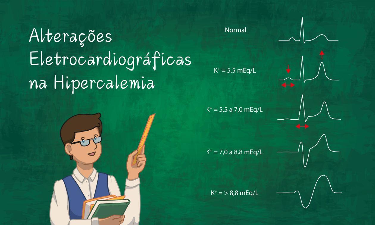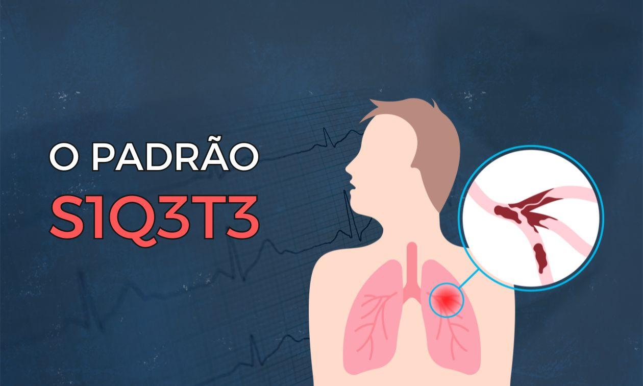

You may have heard that there are unmistakable signs of pulmonary embolism (PE) on the ECG, such as the McGinn-White sign, also known as the S1Q3T3 pattern, a specific finding and therefore supposedly decisive for diagnosing the disease.
But are the ECG in general, and the McGinn-White sign in particular, really that powerful for diagnosing this disease? What are the other classic electrocardiographic signs of PE? How much should the ECG influence our clinical suspicion of this potentially fatal condition?
What Are the Classic Electrocardiographic Signs of PE?
Although the ECG is widely used in the initial evaluation of patients with suspected PE, its diagnostic accuracy is limited. The findings traditionally taught include:
- Tachycardia (HR > 100 bpm)
- Negative or flattened T waves in at least two inferior leads
- R > S in V1
- ST elevation > 1 mm in III
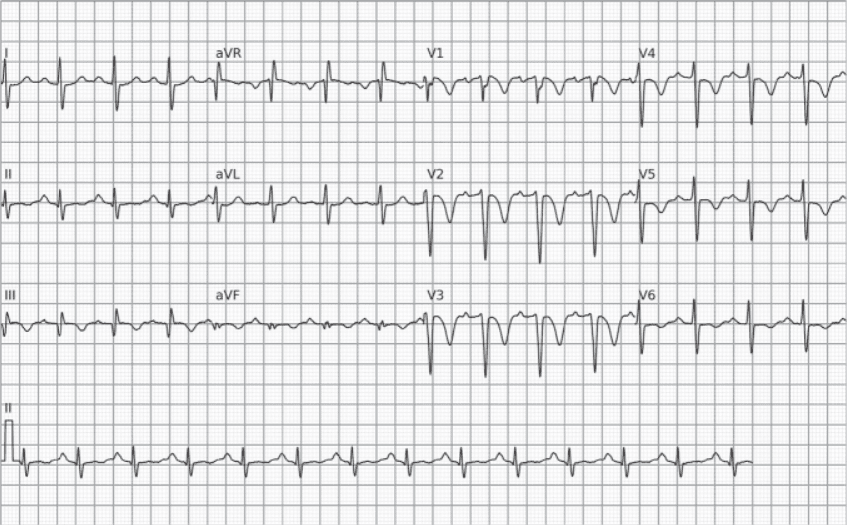
- S1Q3T3 pattern (S wave in I, Q wave and negative T wave in III), with or without a prominent Q wave (≥ 1.5 mm)
- Right bundle branch block (complete or incomplete)
- Right axis deviation (> 90°)
- Right ventricular strain pattern (T-wave inversion or flattening + ST depression ≥ 0.5 mm in at least two inferior leads and two anterior leads from V1 to V3)
Do These Signs Really Help with the Diagnosis?
A new study by Smaniotto, José Alencar and colleagues evaluated 273 patients with suspected PE at a tertiary hospital (Instituto Dante Pazzanese de Cardiologia), comparing ECG findings with the diagnosis confirmed by CT pulmonary angiography (CTPA). The analysis showed that:
- Only tachycardia (OR 1.93) and T-wave inversion in at least two inferior leads (OR 1.82) were independently associated with the diagnosis of PE.
- Despite moderate specificity (85.9%), the classic S1Q3T3 pattern had a positive likelihood ratio (LR+) of only 2.07, indicating marginal clinical usefulness.
- The right ventricular strain pattern, with T-wave inversion or flattening in at least 2 anterior and 2 inferior leads, had an LR+ of 4.75, the most significant of the criteria evaluated, but still of limited diagnostic value.
What Does This Mean in Practice?
Even the “classic” ECG findings should not be used alone to confirm or rule out PE. At best, they increase the diagnostic probability modestly.
Imagine, for example, an elderly man with a history of hypertension and dyslipidemia who arrives at the emergency department with chest pain, dyspnea, hemoptysis and a heart rate of 95 bpm. On initial clinical assessment, his Wells score would be 1 point because of hemoptysis, giving a pretest probability of PE of approximately 1.3%. On the ECG, the only notable finding for PE is the classic S1Q3T3 pattern (LR+ = 2.07). Thus, the post-test probability of the disease after the ECG would rise to only 2.7%, according to the Fagan nomogram below (generated at https://ferramentasmbe.netlify.app/):
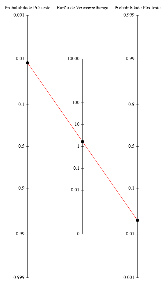
This shows the low clinical relevance of one of the electrocardiographic signs most classically associated with pulmonary embolism, at least in medical school and residency exams.
So Is the ECG Useless in PE?
Not quite. Although it has low diagnostic accuracy, the ECG may have prognostic value. Studies show that certain findings, such as complete right bundle branch block or the strain pattern, are associated with a worse clinical course and right ventricular dysfunction, but they should not be used as a stand-alone diagnostic screening tool.
Conclusion
The ECG remains a low-cost, widely available and useful test in the emergency setting, but it should not be overestimated in the diagnosis of PE. The classic electrocardiographic signs are of marginal value in the workup of this disease. The diagnostic approach should combine structured clinical assessment (such as the Wells score), laboratory markers (such as D-dimer) and imaging, with CT pulmonary angiography as the gold standard for confirmation.
References
- Smaniotto MF, Alencar E Silva GPS, Heringer J de N, Almeida MT de C, Scheffer MK, Felicioni SP, et al. Classical ECG findings in pulmonary embolism have minimal diagnostic accuracy: A cross-sectional study. J Investig Med Off Publ Am Fed Clin Res. June 30, 2025;10815589251357621.
- McGINN S, WHITE PD. ACUTE COR PULMONALE RESULTING FROM PULMONARY EMBOLISM: ITS CLINICAL RECOGNITION. J Am Med Assoc. April 27, 1935;104(17):1473–80.
- Scheffer MK, De Marchi MFN, de Alencar Neto JN, Felicioni SP. Eletrocardiograma de A a Z. São Paulo: Manole, 2024.