

Right bundle branch block (RBBB) is an electrocardiographic abnormality characterized by delayed electrical conduction through the right bundle branch of the His bundle, resulting in late right ventricular depolarization. On the ECG, it classically manifests as an rSR’ pattern in V1 and V2, QRS widening ≥ 120 ms, and secondary repolarization changes in the right precordial leads.
Electrocardiographic Criteria
- QRS duration ≥ 120 ms
- rsr’, rsR’, rSR’, or more rarely qR morphology in V1 and V2. The R’ or r’ wave is usually wider than the initial R wave. In a minority of patients, a broad, notched R wave may be seen in V1 and/or V2
- S wave longer than the R wave or > 40 ms in leads I and V6
Incomplete or First-Degree Right Bundle Branch Block
- According to the American Heart Association guideline, incomplete RBBB is defined by a QRS duration between 110 and 119 ms with the same QRS morphological criteria as “complete” RBBB
- The Brazilian guideline suggests the term terminal conduction delay, emphasizing that it may be a normal variant
ECG in Right Bundle Branch Block
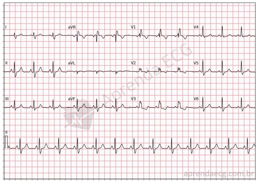
Right bundle branch block. RBBB is characterized by a QRS duration greater than 120 ms and an rsR’ pattern in V1, as well as slurred S waves in leads I, aVL, V5, and V6.
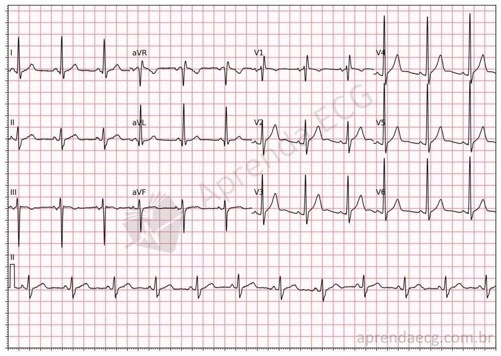
First-degree or partial right bundle branch block. It has the same features as RBBB, but the QRS is not widened and its duration is between 110 and 119 ms.
Related Topics
References
- Scheffer MK, De Marchi MFN, de Alencar Neto JN, Felicioni SP. Eletrocardiograma de A a Z. São Paulo: Manole, 2024.
- Surawicz B, Childers R, Deal BJ, Gettes LS. AHA/ACCF/HRS Recommendations for the Standardization and Interpretation of the Electrocardiogram. Part III: Intraventricular Conduction Disturbances. J Am Coll Cardiol. 2009;53(11):996–981.
- Samesima N, God EG, Kruse JCL, Leal MG, et al. Diretriz da Sociedade Brasileira de Cardiologia sobre a Análise e Emissão de Laudos Eletrocardiográficos – 2022. Arq Bras Cardiol. 2022;93(3 Suppl 2):2–19.


