

Left bundle branch block (LBBB) is an electrocardiographic abnormality that reflects delayed or interrupted electrical conduction through the left bundle branch of the His bundle, resulting in abnormal and delayed left ventricular depolarization. On the ECG, it is characterized by QRS widening ≥ 120 ms, absence of Q waves in the lateral leads (I, V5, and V6), and an rS or QS pattern in V1 and V2.
Electrocardiographic Criteria
- Wide QRS with duration ≥ 120 ms
- Absence of “q” waves in I, aVL, V5, and V6; variants may have a “q” wave in aVL only
- Broad R waves with mid-to-late notching and/or slurring in I, aVL, V5, and V6
- “r” wave with slow progression from V1 to V3; QS complexes may occur
- Intrinsicoid deflection in V5 and V6 ≥ 50 ms
- QRS electrical axis between -30° and +60°
- Asymmetric ST depression and T wave opposite to the mid-to-late conduction delay
Strauss proposed stricter criteria for the electrocardiographic diagnosis of LBBB, which include: (1) QRS duration > 140 ms (men) or > 130 ms (women), (2) QS or rS in V1 and V2, and (3) mid-QRS notching or slurring in 2 of the leads V1, V2, V5, V6, I, and aVL.
ECG in Left Bundle Branch Block
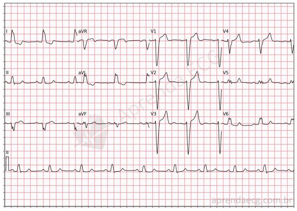
Left bundle branch block. In LBBB, the QRS complex lasts longer than 120 ms and is negative in V1, with an rS or QS morphology. In leads I, aVL, V5, and V6, the QRS shows mid-to-late notching or slurring with a “tower-like” appearance.
Further Reading
Related Topics
References
- Scheffer MK, De Marchi MFN, de Alencar Neto JN, Felicioni SP. Eletrocardiograma de A a Z. São Paulo: Manole, 2024.
- Samesima N, God EG, Kruse JCL, Leal MG, et al. Diretriz da Sociedade Brasileira de Cardiologia sobre a Análise e Emissão de Laudos Eletrocardiográficos – 2022. Arq Bras Cardiol. 2022;93(3 Suppl 2):2–19.
- Strauss DG, Selvester RH, Wagner GS. Defining left bundle branch block in the era of cardiac resynchronization therapy. Am J Cardiol. 2011;107(6):927–34.


