

Left posterior fascicular block (LPFB) is an electrocardiographic finding caused by delayed or blocked conduction through the posteroinferior fascicle of the left bundle branch of the His bundle. This block shifts the heart's electrical axis to the right, usually between +90° and +180°, with normal or slightly widened QRS complexes (<120 ms). An rS pattern is seen in leads I and aVL and a qR pattern in II, III, and aVF. It is important to note that the posteroinferior fascicle is the least vulnerable part of the intraventricular conduction system; therefore, isolated LPFB is extremely rare.
Electrocardiographic Criteria
- Frontal plane axis deviation between 90° and 180°
- rS pattern in I and aVL
- qR pattern in II, III, and aVF, with the R wave in III > R wave in II
- Ventricular activation time > 45 ms in aVF
- QRS duration < 120 ms
These criteria are valid only in the absence of a slender (asthenic) body habitus, RVH, or a lateral electrically inactive area.
ECG in Left Posterior Fascicular Block
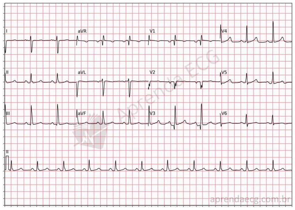
Left posterior fascicular block. Isolated LPFB is rare; it is more commonly associated with RBBB. In this example, note the right axis deviation and the rS morphology in I and aVL and qR in II, III, and aVF (with the R wave in III taller than the R wave in II).
Related Topics
References
- Surawicz B, Childers R, Deal BJ, Gettes LS. AHA/ACCF/HRS Recommendations for the Standardization and Interpretation of the Electrocardiogram. Part III: Intraventricular Conduction Disturbances. J Am Coll Cardiol. 2009;53(11):996–981.
- Samesima N, God EG, Kruse JCL, Leal MG, et al. Diretriz da Sociedade Brasileira de Cardiologia sobre a Análise e Emissão de Laudos Eletrocardiográficos – 2022. Arq Bras Cardiol. 2022;93(3 Suppl 2):2–19.
- Elizari M V. Fascicular Blocks: Update 2019. Curr Cardiol Rev. 2021;17(1):31–40.


