Left bundle branch block (LBBB) is an electrocardiographic abnormality characterized by delayed electrical conduction through the left bundle branch of the His bundle. This condition results in abnormal ventricular activation, significantly altering the normal sequence of cardiac depolarization. LBBB may be associated with various structural heart diseases and has important prognostic impact.
Mechanism of Left Bundle Branch Block
In LBBB, the electrical impulse is conducted normally through the right bundle branch, depolarizing the right ventricle first. Depolarization of the left ventricle occurs in a retrograde, delayed fashion through the ventricular myocardium. This produces a widened QRS complex and characteristic changes on the electrocardiogram.
Ventricular activation in LBBB occurs in three main phases:
- Initial activation through the right bundle branch: occurs normally, without delay.
- Transseptal depolarization: occurs from right to left, altering QRS morphology.
- Late depolarization of the left ventricle: results in broad, notched R waves in the lateral leads.
Electrocardiographic Criteria for the Diagnosis of LBBB
The classic criteria for the diagnosis of LBBB include:
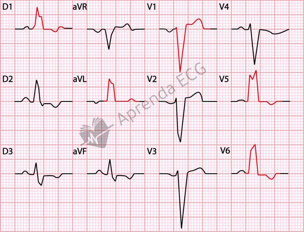
- QRS duration ≥ 120 ms
- Absence of a “q” wave in I, aVL, V5 and V6 (some variants may show a “q” wave in aVL)
- Broad R waves with notching or slurring in I, aVL, V5 and V6
- Slow progression of “r” waves from V1 to V3, with a possible QS pattern
- Intrinsicoid deflection ≥ 50 ms in V5 and V6
- QRS axis between -30° and +60°
- ST-segment depression and T-wave inversion opposite to the mid-terminal delay
The Strauss criteria propose a stricter diagnosis of LBBB, including:
- QRS > 140 ms in men and > 130 ms in women
- QS or rS in V1 and V2
- Mid-QRS notching or slurring in at least two leads (V1, V2, V5, V6, I and aVL)
Clinical and Electrocardiographic Considerations
- LBBB rarely occurs in structurally normal hearts, and its presence should prompt investigation for underlying heart disease.
- Its prevalence in the general population is below 1% and increases with age.
- The prognosis of LBBB varies according to the associated heart disease.
- When present in patients with coronary artery disease or heart failure, LBBB is an independent predictor of mortality.
- In asymptomatic patients without structural heart disease, the prognostic impact of LBBB remains controversial.
- Cardiac resynchronization therapy (CRT) may benefit symptomatic patients with heart failure and reduced ejection fraction, especially if the QRS is ≥ 150 ms with LBBB morphology.
Etiologies of Left Bundle Branch Block
LBBB can be caused by several conditions, including:
- Cardiomyopathies
- Valvular heart disease (especially aortic stenosis)
- Coronary artery disease and myocardial ischemia
- Hypertension
- Myocarditis
- Degenerative disease of the conduction system (Lev-Lenègre disease)
- Cardiac surgical procedures, such as transcatheter aortic valve implantation (TAVI)
Conclusion
Left bundle branch block is an important electrocardiographic abnormality with variable clinical impact. Its presence requires a detailed evaluation to identify underlying heart disease, and recognizing it is essential for risk stratification and clinical management.
References
- Scheffer MK, De Marchi MFN, de Alencar Neto JN, Felicioni SP. Eletrocardiograma de A a Z. São Paulo: Manole, 2024.
- Samesima N, God EG, Kruse JCL, Leal MG, et al. Diretriz da Sociedade Brasileira de Cardiologia sobre a Análise e Emissão de Laudos Eletrocardiográficos – 2022. Arq Bras Cardiol. 2022;93(3 Suppl 2):2–19.
- Strauss DG, Selvester RH, Wagner GS. Defining left bundle branch block in the era of cardiac resynchronization therapy. Am J Cardiol. 2011;107(6):927–34.