Fascicular blocks are one of the most controversial topics in the current electrocardiographic literature. These discussions begin at the foundation of medicine: anatomy. Discrepancies in how the fascicles of the right and left bundle branches are described create uncertainty about the nomenclature and criteria for interpreting these blocks on the ECG, since the classifications depend directly on how these fascicles are organized.
Anatomy
The anatomy of the terminal portion of the right and left bundle branches is the subject of different theories. The trifascicular theory describes two fascicles in the left bundle branch (anterosuperior and posteroinferior) and a right bundle branch that gives off no fascicles. On the other hand, the tetrafascicular theory, the most widely accepted in the literature, proposes a third fascicle in the left bundle branch: the anteromedial (septal) fascicle. This is the theory we will use here to define fascicular blocks. There is also the hexafascicular theory, which proposes three additional fascicles in the right bundle branch but still lacks broad acceptance in the scientific community.
Relevance to the ECG
Fascicular blocks result from interruption of electrical conduction in the fascicles of the His bundle. According to the tetrafascicular theory, block of the anterosuperior fascicle produces left anterior fascicular block (LAFB), while block of the posteroinferior fascicle causes left posterior fascicular block (LPFB). Likewise, block of the anteromedial fascicle is associated with left septal fascicular block (LSFB), although its electrocardiographic criteria are still debated. Because the tetrafascicular theory does not recognize fascicles in the right bundle branch, there is no classification of fascicular blocks for that branch.
Controversies
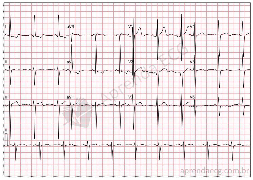
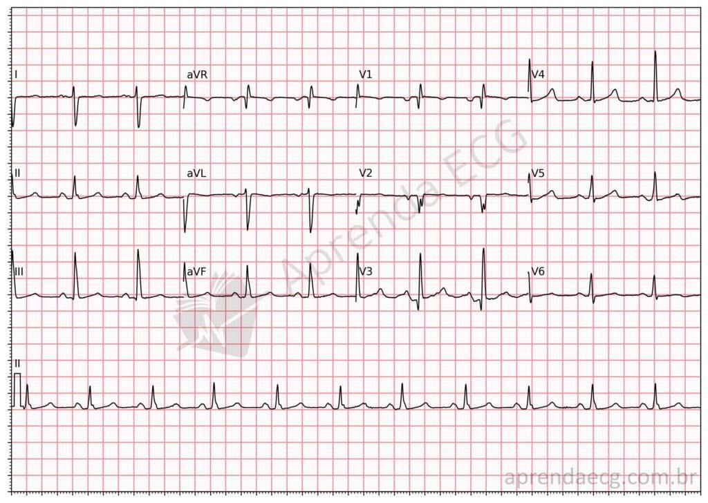
Although the tetrafascicular theory is the most widely accepted, some publications, such as the 2022 Brazilian Guideline on the Analysis and Reporting of Electrocardiograms, adopt the concept of “right bundle branch fascicular blocks”, based on the hexafascicular theory. However, this theory lacks a sufficiently solid foundation to support the classification of fascicular blocks, because including additional fascicles in the right bundle branch creates diagnostic uncertainty and compromises the uniformity and accuracy of ECG interpretation. For this reason, the tetrafascicular theory remains the most robust and reliable model for classifying fascicular blocks.
Conclusion
Understanding the anatomy of the fascicles is essential for proper classification of bundle branch blocks. The tetrafascicular theory offers the most consistent model for interpreting fascicular blocks, dividing them into LAFB, LPFB, and LSFB. This nomenclature reflects the anatomical organization of the left bundle branch and avoids ambiguities such as distinguishing a “left” anterosuperior fascicular block from a “right” one, since there is no corresponding fascicle in the right bundle branch. Therefore, any attempt to define “right bundle branch fascicular blocks” lacks anatomical and scientific support.
Learn more
Visit our ECG Library and review the electrocardiographic criteria for LAFB, LPFB and LSFB. Learn to recognize these patterns on the tracing and enrich your clinical practice.
References
- Alencar JN. Tratado de ECG. 2022.
- Scheffer MK, De Marchi MFN, de Alencar Neto JN, Felicioni SP. Eletrocardiograma de A a Z. São Paulo: Manole, 2024.
- de Alencar Neto JN. Letter to editor: Right fascicles: A call for empirical evidence. J Electrocardiol. 2024;83:118.
- Samesima N, God EG, Kruse JCL, Leal MG, et al. Diretriz da Sociedade Brasileira de Cardiologia sobre a Análise e Emissão de Laudos Eletrocardiográficos – 2022. Arq Bras Cardiol. 2022;93(3 Suppl 2):2–19.