Today we will learn how to identify right bundle branch block (RBBB) on the electrocardiogram (ECG) in a simple, practical way. RBBB is an electrocardiographic abnormality characterized by delayed electrical conduction through the right bundle branch of the His bundle. This delay produces a specific ECG pattern that can be easily identified once we understand its mechanism and electrocardiographic criteria.
Understanding the Mechanism of Right Bundle Branch Block
In the healthy heart, the electrical impulse travels rapidly through the conduction system via the right and left bundle branches, allowing simultaneous depolarization of the ventricles. In RBBB, the right bundle branch has a block that prevents or delays this electrical conduction.
How does this affect the ECG?
- Preserved initial activation:
- The left bundle branch conducts the impulse normally, depolarizing first the interventricular septum and then the left ventricle.
- This initial activation is not altered in RBBB.
- Right ventricular depolarization through alternative routes:
- The electrical impulse reaches the right ventricle indirectly, through the ventricular muscle, via the interventricular septum and the left ventricular myocardium. This process is slower and produces QRS widening (≥ 120 ms).
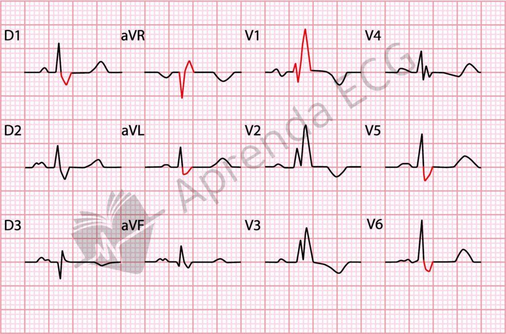
- In V1, you will see an additional R’ wave (remember: an extra R wave at the end of the QRS complex). This morphology is called rsr’, rsR’, or rSR’, depending on its exact shape. This R’ represents the delayed activation of the right ventricle, since the block prevents the impulse from traveling normally down the right bundle branch.
- In the Lateral Leads (V5, V6, I, aVL), look for a slurred S wave, that is, a longer S wave reflecting the terminal delay in ventricular depolarization.
- Secondary repolarization changes:
- Delayed right ventricular depolarization affects the ventricular repolarization pattern, resulting in a negative T wave in the right precordial leads (V1 and V2).
Look at the figure below and identify the changes caused by RBBB (highlighted in red).
Incomplete or First-Degree RBBB
What if the QRS is widened but does not reach 120 ms? In that case, we have what is called incomplete or first-degree RBBB. The general features are the same as in complete RBBB, but the QRS duration is shorter, between 110 and 120 ms.
Incomplete RBBB is more common in healthy individuals and does not always have clinical significance.
Summary: How to Recognize RBBB on the ECG
- QRS duration: widened (> 120 ms in complete RBBB; 110–120 ms in incomplete RBBB).
- V1: presence of an R’ wave (rsr’, rsR’, or rSR’), representing delayed right ventricular depolarization.
- V5, V6, I, aVL: slurred, prolonged S wave.
- T wave: negative in the right-sided leads (V1 and V2), as a result of altered ventricular repolarization.
Why Is RBBB Important?
Although isolated RBBB may be benign, it can also be a sign of underlying heart disease, such as myocarditis, pulmonary hypertension, or pulmonary embolism. Recognizing these patterns on the ECG is essential for the diagnosis and management of more serious conditions.
Now that you know how to identify RBBB, practice by analyzing real ECGs and comparing patterns. This will help consolidate what you have learned and make sure you are ready to recognize RBBB confidently in daily clinical practice.
References
- Scheffer MK, De Marchi MFN, de Alencar Neto JN, Felicioni SP. Eletrocardiograma de A a Z. São Paulo: Manole, 2024.
- Surawicz B, Childers R, Deal BJ, Gettes LS. AHA/ACCF/HRS Recommendations for the Standardization and Interpretation of the Electrocardiogram. Part III: Intraventricular Conduction Disturbances. J Am Coll Cardiol. 2009;53(11):996–981.