In the daily routine of the emergency department, most electrocardiograms are reassuring. Some, however, allow no hesitation. These are patterns the on-call physician simply cannot miss.
We are not always facing classic ST-segment elevation. In many cases, the risk lies in the QRS morphology, the ventricular rate, dissociation between the atria and ventricles, or changes that mimic infarction but reflect impending systemic collapse.
In these settings, the ECG works as an early marker of electrical instability and prognosis. Recognizing these patterns in the first few minutes may mean activating the cath lab, performing cardioversion, starting circulatory support or rapidly investigating a critical non-cardiac condition.
Below, we discuss five high-risk electrocardiographic patterns that every emergency physician and cardiologist should recognize immediately.
1. Shark Fin Sign
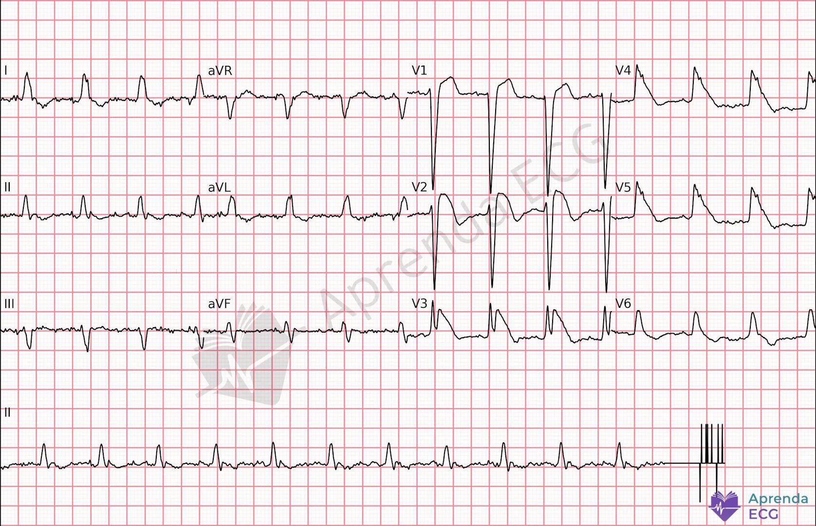
The so-called shark fin sign, also described as the lambda wave or triangular QRS-ST-T pattern, is one of the most threatening patterns in acute coronary syndrome.
How to recognize it
- Fusion of the QRS complex, ST segment and T wave
- Giant R wave (≥ 10 mm)
- Broad triangular morphology
- Disappearance of the ST segment as a separate structure
Visually, the tracing resembles a “fin” or the Greek letter lambda.
What it means
This pattern is associated with:
- Proximal occlusion of the left anterior descending artery (LAD)
- Proximal occlusion of the right coronary artery
- Absence of collateral circulation
Studies show significantly higher mortality with lambda/triangular patterns compared with typical ST elevation, with a higher incidence of cardiogenic shock and VF.
Practical implication
- Immediate activation of a reperfusion strategy
- Intensive monitoring
- Low threshold for circulatory support
2. Pre-excited Atrial Fibrillation
Pre-excited AF is an electrical emergency, regardless of the clinical context in which it occurs.
How to recognize it
- Irregularly irregular rhythm
- Absence of organized P waves
- Wide, pleomorphic QRS complexes
- Beat-to-beat variation in QRS morphology
- Ventricular rate often > 200 bpm
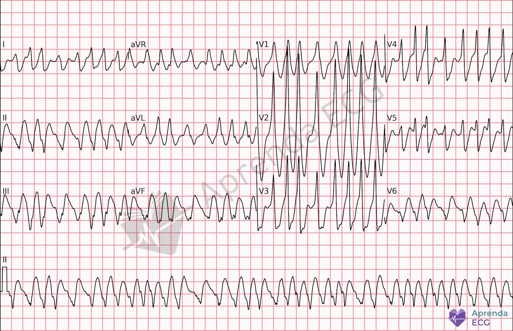
The QRS changes because some impulses are conducted through the AV node and some through the accessory pathway.
The real risk
If the accessory pathway has a short refractory period:
- Extremely rapid ventricular conduction
- Degeneration into ventricular fibrillation
- Hemodynamic collapse
Classic mistake
Use of AV nodal blocking agents (adenosine, verapamil, digoxin).
Blocking the AV node favors conduction exclusively over the accessory pathway.
Management
- Synchronized electrical cardioversion if unstable
- Avoid AV nodal blocking agents
3. Complete Atrioventricular Block (Complete Heart Block)
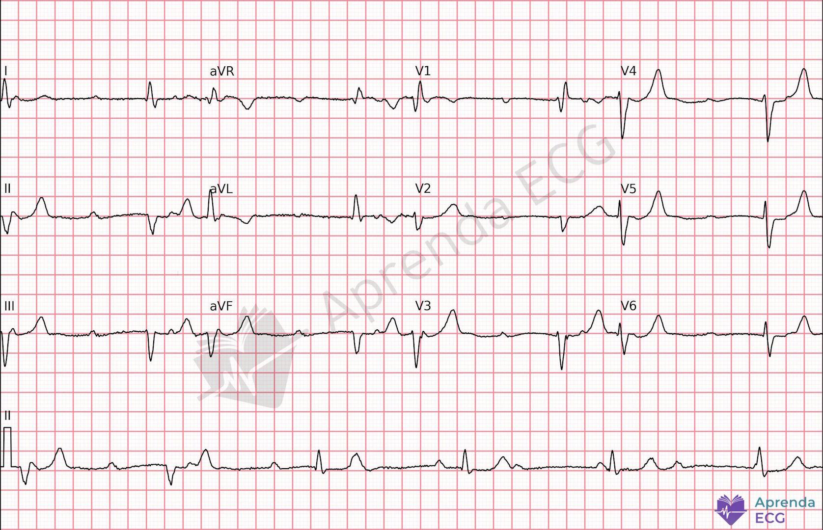
Third-degree atrioventricular block (complete heart block) is characterized by complete AV dissociation.
How to recognize it
- P waves with no fixed relationship to the QRS
- Atrial rate higher than the ventricular rate
- Regular escape rhythm
- Narrow QRS (junctional escape) or wide QRS (ventricular escape)
Etiologies and clinical context
Complete heart block can occur in multiple settings:
- Acute myocardial infarction (especially anterior or inferior)
- Degeneration of the His-Purkinje system
- Myocarditis
- After cardiac surgery
- Drug toxicity
When the block is infranodal (wide QRS, escape < 40 bpm), the risk of instability is higher, regardless of the cause.
Practical implication
- Immediate transcutaneous pacing if unstable
- Consider transvenous pacemaker
- Continuous monitoring
Not every complete heart block carries the same prognostic weight. The pattern of the ventricular escape helps estimate severity and urgency.
4. Type 1 Brugada Pattern
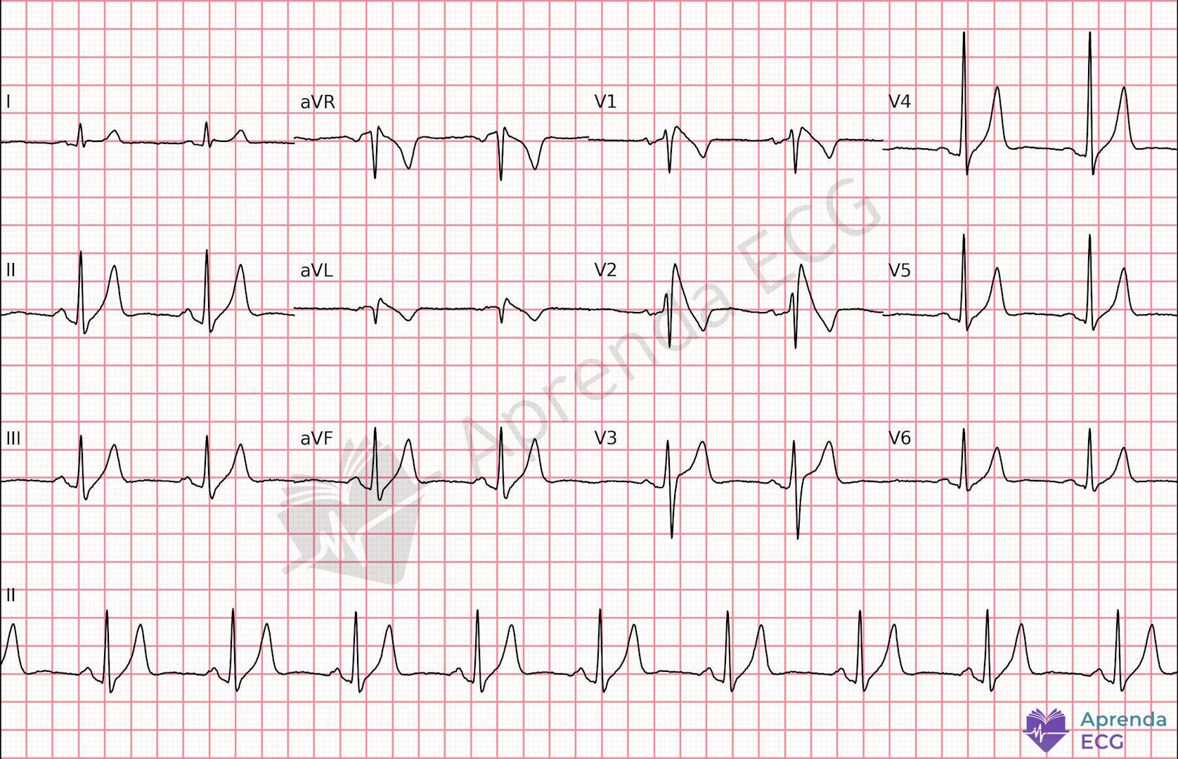
The type 1 Brugada pattern can be unmasked by fever, metabolic disturbances and also by ischemia.
How to recognize it
- ST elevation ≥ 2 mm in V1–V3
- Coved morphology (“coved type”)
- Symmetric negative T wave
Why it matters
- Increased risk of polymorphic VT and ventricular fibrillation
- Can be unmasked by fever, metabolic disturbances or drugs
- May coexist with structural heart disease, but it is primarily a channelopathy
Practical implication
- Differentiate from classic ischemic ST elevation
- Continuous monitoring
- Evaluation for arrhythmic risk stratification
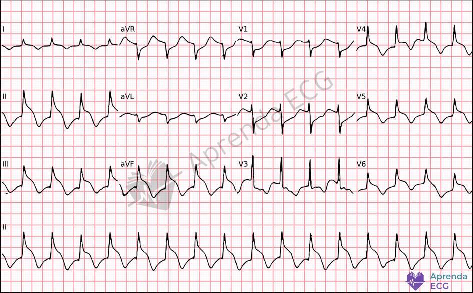
5. Spiked Helmet Sign
The spiked helmet sign is a marker of critical systemic illness with high mortality.
How to recognize it
- ST-segment elevation with baseline shift
- Marked QT prolongation (often > 550 ms)
- Dome-shaped morphology with a spike resembling a Prussian helmet
Typical clinical context
- Sepsis
- Intracranial hemorrhage
- Severe metabolic disturbances
In published series, in-hospital mortality exceeds 50%.
Practical implication
- Look for a critical extracardiac cause
- Monitor for the risk of torsades de pointes
Conclusion
Recognizing high-risk ECG patterns takes more than measuring millimeters at the J point.
Some tracings indicate:
- Impending electrical instability
- Cardiogenic shock
- Risk of sudden death
The ECG remains the fastest tool for initial risk stratification. Knowing how to identify these five patterns can change management in the first few minutes and, in many cases, prevent abrupt deterioration or sudden death.
References
- Scheffer MK, De Marchi MFN, de Alencar Neto JN, Felicioni SP. Eletrocardiograma de A a Z. São Paulo: Manole; 2024.