The ECG in the Diagnosis of Hypertrophic Cardiomyopathy (HCM)
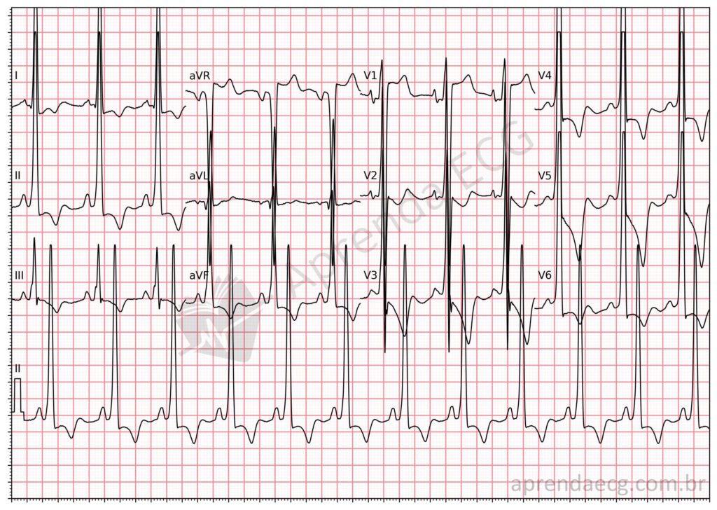
The ECG is often the first test performed in patients with suspected HCM and may be the only manifestation of the disease in its early stages. Although there is no pathognomonic electrocardiographic pattern for HCM, the ECG shows some type of abnormality in more than 90% of patients. Taken together, these abnormalities raise diagnostic suspicion.
- Atrial Abnormalities: Left atrial enlargement, reflecting diastolic dysfunction and increased filling pressures, is common in HCM and may be evident on the ECG. However, these abnormalities rarely appear in isolation in HCM
- Ventricular Pre-excitation: Ventricular pre-excitation with marked left ventricular hypertrophy may suggest an association with glycogen storage diseases, such as those caused by mutations in the LAMP2 or PRKAG2 genes.
- Q Waves: Deep, narrow Q waves (> 25% of the R wave and < 30 ms in duration) in the inferolateral leads, called “dagger-like” Q waves, are among the features of the disease. When accompanied by tall R waves in leads V1 to V3, they raise a strong suspicion of HCM, depending on the clinical context.
- QRS Complex and Ventricular Hypertrophy: Increased QRS voltage, such as meeting the Cornell and Sokolow-Lyon criteria, is a common finding. However, only 2% of patients with HCM have increased QRS voltage without other ECG abnormalities. Bundle branch blocks are rare in HCM and may appear after invasive procedures, such as septal myectomy.
- Repolarization Abnormalities: Deep negative T waves (> 2 mm), usually preceded by ST-segment depression in the inferolateral leads, are very frequent findings in HCM. So-called “giant T waves” (inverted T waves > 10 mm in amplitude) in the precordial leads are characteristic of apical HCM (Yamaguchi disease).
- QTc Prolongation: QTc prolongation (> 480 ms) occurs in 1 in 8 patients with HCM and seems to reflect electrophysiological remodeling of the cardiomyocytes.
ECG and Risk Stratification in HCM
Some electrocardiographic parameters have been shown to be associated with the prognosis of the disease:
- QRS Duration: A wide QRS (>120ms) has been associated with a higher risk of sudden death and ICD shocks.
- Low QRS Voltage: Low voltage in the frontal plane may indicate extensive myocardial fibrosis and is associated with a worse prognosis.
- “Pseudo-Infarction” Pattern: ST-segment elevation in the precordial leads has been linked to a higher risk of adverse events.
- Pathological T-Wave Inversion: Deep T-wave inversions, especially in the precordial leads, may indicate a higher risk of ventricular arrhythmias.
- ST-Segment Depression: ST-segment depression > 2mm may also be associated with higher risk.
- QTc Interval: QTc prolongation (> 480ms) has been associated with a higher risk of appropriate ICD shocks
- Necrosis Q Waves: Q waves lasting > 30 ms indicate extensive fibrosis and are associated with a higher risk of events.
ECG and Differential Diagnoses in HCM:
The ECG plays a crucial role in the differential diagnosis between sarcomeric HCM and its phenocopies, as well as in distinguishing the “athlete's heart” from normal variants.
Sarcomeric HCM vs. Phenocopies
Deep Q waves with positive T waves in the same leads, or giant T-wave inversions in the anterolateral leads, suggest sarcomeric HCM, whereas a short PR interval and ventricular pre-excitation are more common in storage diseases. Low QRS voltage suggests cardiac amyloidosis.
“Athlete's Heart” vs. HCM
The ECG can help differentiate the physiological adaptations of the athlete's heart from pathological HCM. Isolated voltage criteria for left ventricular hypertrophy or left axis deviation of the QRS suggest normal adaptation to exercise. Repolarization abnormalities, such as T-wave inversion in the lateral leads, ST-segment depression, and pathological Q waves, are more suggestive of HCM and require further investigation.
References
- Finocchiaro G, Sheikh N, Biagini E, Papadakis M, Maurizi N, Sinagra G, et al. The electrocardiogram in the diagnosis and management of patients with hypertrophic cardiomyopathy. Hear Rhythm [Internet]. 2020;17(1):142–51.
- Gossios T, Savvatis K, Zegkos T, Ntelios D, Rouskas P, Parcharidou D, et al. Deciphering hypertrophic cardiomyopathy with electrocardiography. Heart Fail Rev [Internet]. 2022;27(4):1313–23.
- Scheffer MK, De Marchi MFN, de Alencar Neto JN, Felicioni SP. Eletrocardiograma de A a Z. São Paulo: Manole, 2024.