A tall R wave in leads V1 and V2, defined as an R/S ratio > 1 or a pure R wave, is a finding that should always draw attention. Although it may be seen as a normal variant (especially in children or in adults with counterclockwise rotation of the heart), identifying it requires careful analysis, as it may indicate structural heart disease or technical errors.
In this article, we review the 10 main causes of a prominent R wave in V1 and V2, focusing on the electrophysiological mechanisms and the most relevant diagnostic clues.
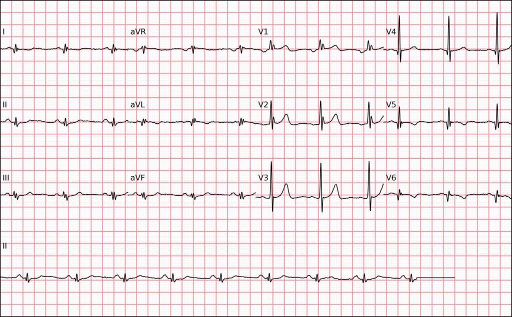
1. 🧱 Electrically Inactive Area in the Lateral Wall
Fibrosis or necrosis in the lateral wall of the left ventricle (as in old infarctions) can produce a tall R wave in V1/V2, a phenomenon known as the “mirror image” of the Q waves in the lateral leads (V5–V6 or V7–V9). In these cases, repolarization changes with an upright T wave in V1 are also often seen.
🧪 Diagnostic tip: record the posterior leads (V7–V9) and look for a lateral scar on cardiac MRI or a history of prior infarction.
2. 🔵 Right Bundle Branch Block (RBBB)
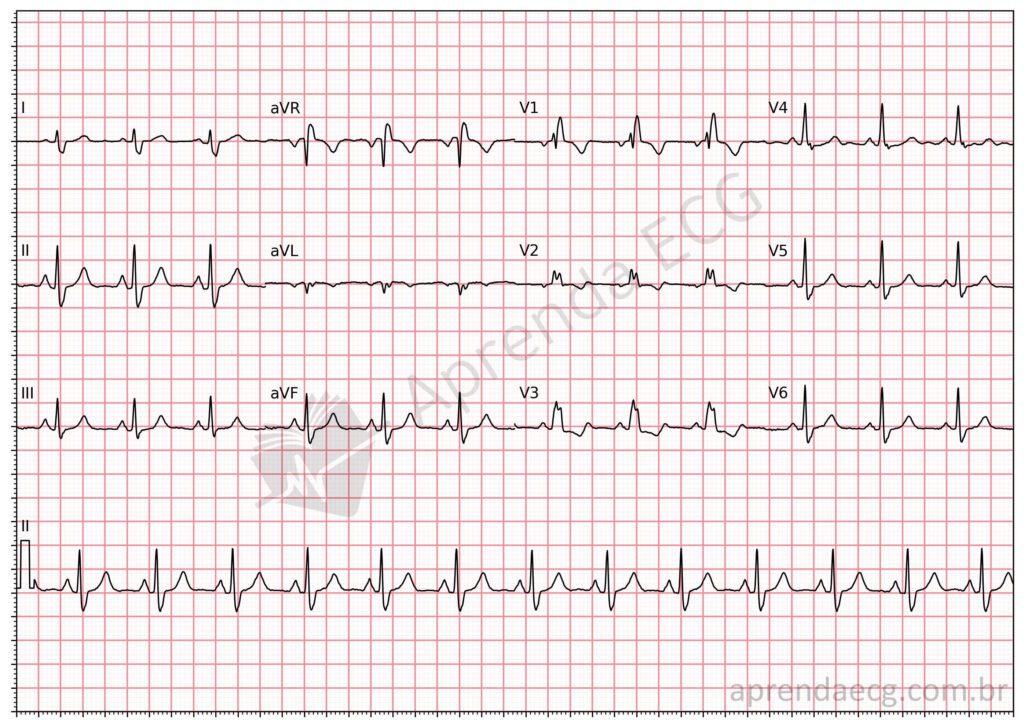
Complete RBBB is characterized by QRS ≥ 120 ms and an rSR’ pattern or pure R wave in V1. Here, the tall R wave reflects delayed activation of the right ventricle.
🧪 Associated clues: terminal QRS slurring in V6 and I, normal axis, and a slurred S wave.
3. 📐 Left septal fascicular block
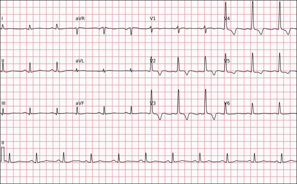
This block affects an anteromedial fascicle of the left bundle branch, redirecting the initial QRS vector anteriorly. The result may be an R wave that becomes progressively taller from V1 to V3.
🧪 Important: it is a diagnosis of exclusion, and its recognition is still controversial.
4. 🧬 Hypertrophic Cardiomyopathy (HCM)
In the asymmetric septal form of HCM, hypertrophy of the interventricular septum can produce tall R waves in V1 and V2. The ECG may also show signs of left ventricular hypertrophy and inferolateral Q waves.
🧪 Associated with: family history, unexplained syncope, and an increased risk of sudden death.
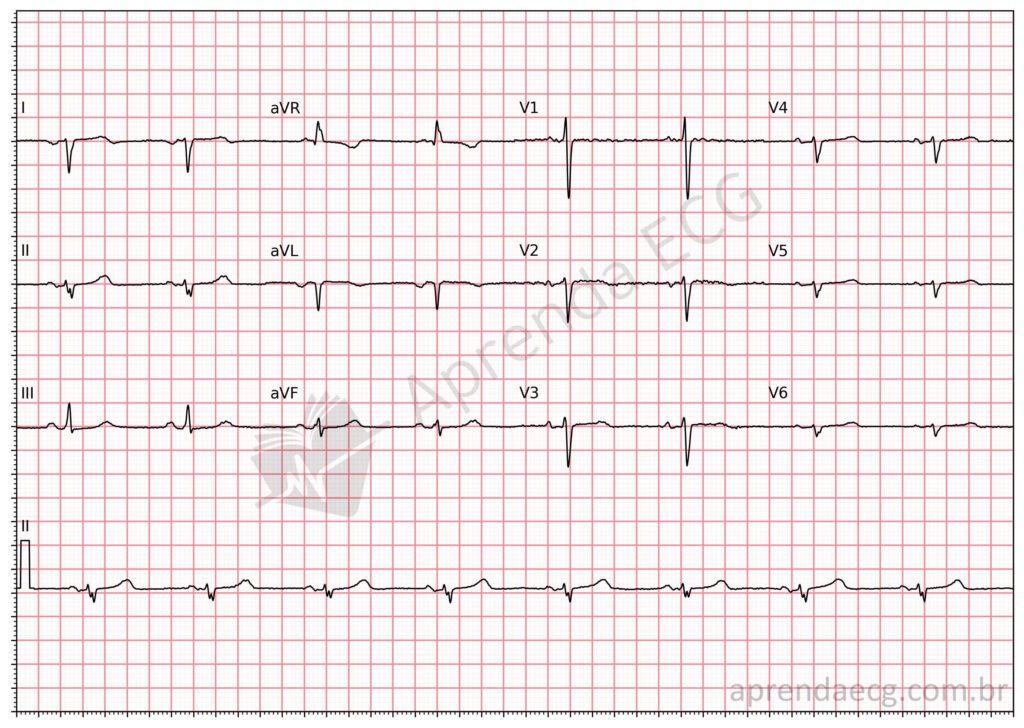
5. 🔄 Dextrocardia
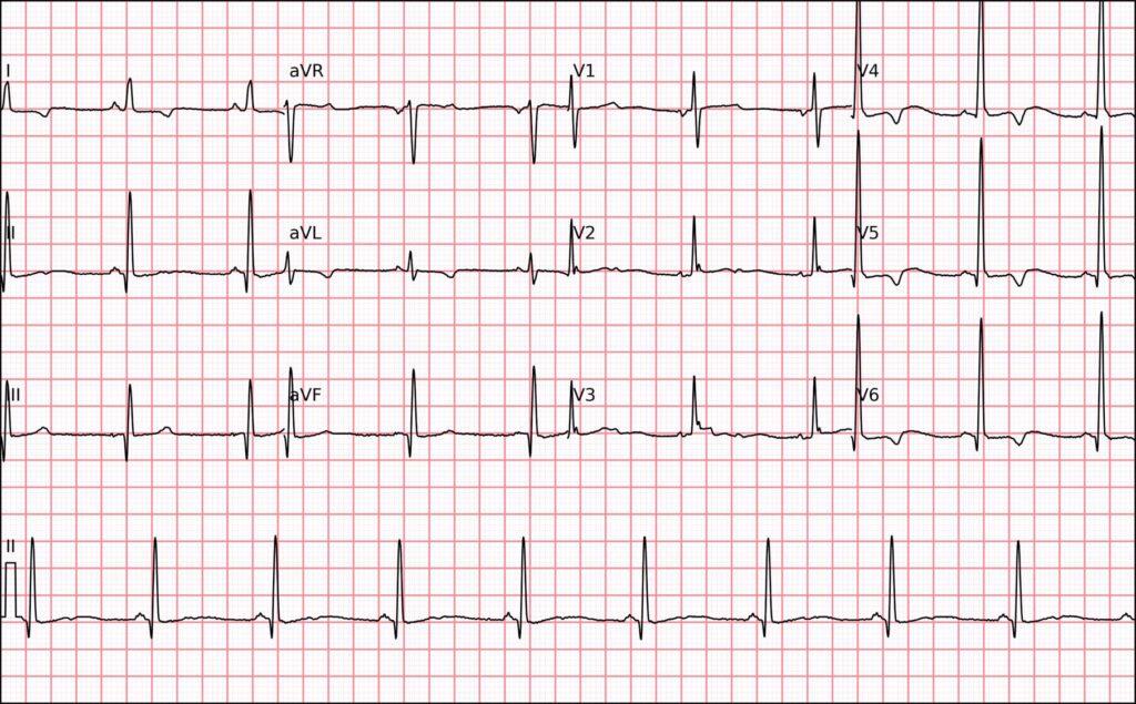
In dextrocardia, the heart points toward the right side of the chest. With the electrodes placed on the left, V1 and V2 record the LV vectors “from behind”, which can produce a tall R wave.
🧪 Diagnostic clues: negative P wave in I, right axis deviation, and low voltage from V3 to V6.
6. 💪 Duchenne Muscular Dystrophy
In this X-linked genetic disease, fibrosis of the LV inferolateral wall can produce tall R waves in V1/V2 and deep Q waves in V5-V6.
🧪 Important: the ECG may be an early marker of myocardial involvement, even before overt systolic dysfunction.
7. ⚠️ Precordial Electrode Misplacement
A common technical error, especially V1/V2 reversal or cranial displacement of the electrodes.
🧪 Suggestion: repeat the ECG, carefully checking the anatomical position of the electrodes.
8. ⚡ Ventricular Preexcitation
When the accessory pathway is left-sided, early ventricular activation travels from left to right, producing a delta wave that contributes to the tall R wave in V1.
🧪 Other findings: short PR, QRS widening, and secondary ventricular repolarization abnormalities.
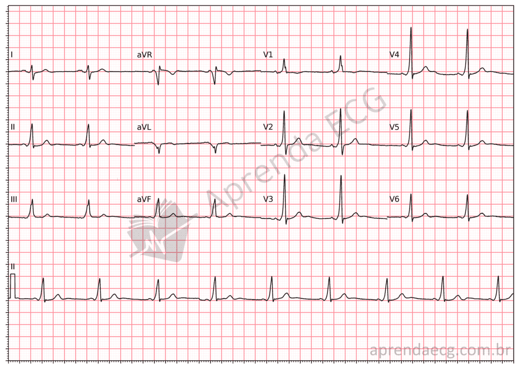
9. ➡️ Right Ventricular Hypertrophy (RVH)
Conditions such as pulmonary hypertension, pulmonary embolism, or congenital heart disease can lead to RVH with a qR pattern or pure R wave in V1, along with right axis deviation.
🧪 Other signs: deep S waves in V5-V6 and right atrial enlargement (P ≥ 2.5 mm in II).
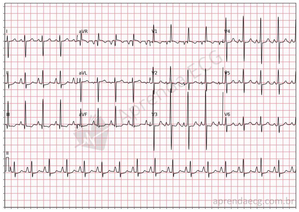
10. ✅ Normal Variant
Counterclockwise rotation of the heart can place the septum more perpendicular to V1, producing larger R waves. In this setting, the finding is isolated, without other pathological changes.
🧪 Important: it should be considered a diagnosis of exclusion after structural and technical causes have been ruled out.
📚 Conclusion
A prominent R wave in V1 and V2 should not be considered a benign finding without careful analysis. Its interpretation depends on the clinical context, the presence of other ECG abnormalities, and, when necessary, confirmation by imaging.
Correctly identifying its cause can reveal diseases such as HCM, muscular dystrophies, or congenital heart disease and, at the same time, avoid misdiagnosis in cases of anatomical variants or technical errors.
References
- Scheffer MK, De Marchi MFN, de Alencar Neto JN, Felicioni SP. Eletrocardiograma de A a Z. São Paulo: Manole, 2024.