Junctional rhythm arises in the atrioventricular (AV) junction, which extends from the input pathways of the AV node to the bifurcation of the His bundle; because the surface ECG rarely allows the focus to be localized, "junctional rhythm" replaced the older "nodal rhythm."1,2 It appears in healthy young people during sleep, in complete atrioventricular block, in digitalis toxicity, and after cardiac surgery; the tracing looks similar, but the meaning does not.1,3
Mechanism
- Intrinsic rate: 40 to 60 bpm in adults; usually higher in children.1,3,4
- Passive (escape or backup): the sinus node becomes slower than the junction, or the sinus impulse does not reach the ventricles, and the junction takes over at its own rate.1,3
- Active: junctional automaticity increases and exceeds the sinus rate, without sinus node failure.1,3
Criteria
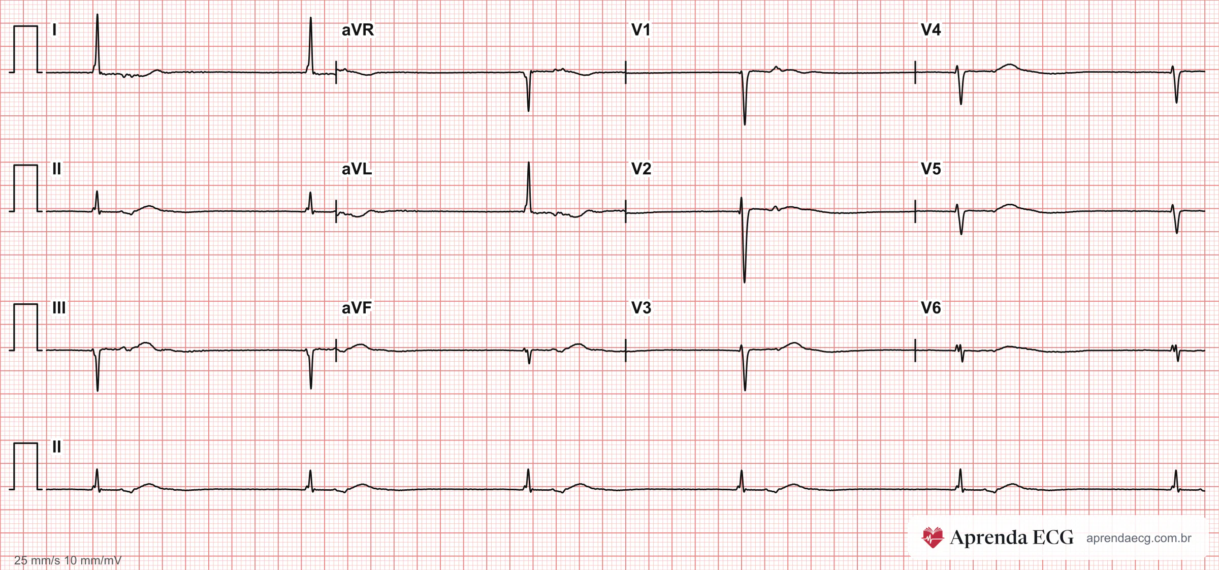
- QRS: identical to or slightly different from baseline, because the ventricles are activated through the His-Purkinje system; regular RR in escape rhythm.1,4,5
- Retrograde P wave: negative in leads II, III, and aVF and positive in aVR, usually narrower than the sinus P wave.1,2,4
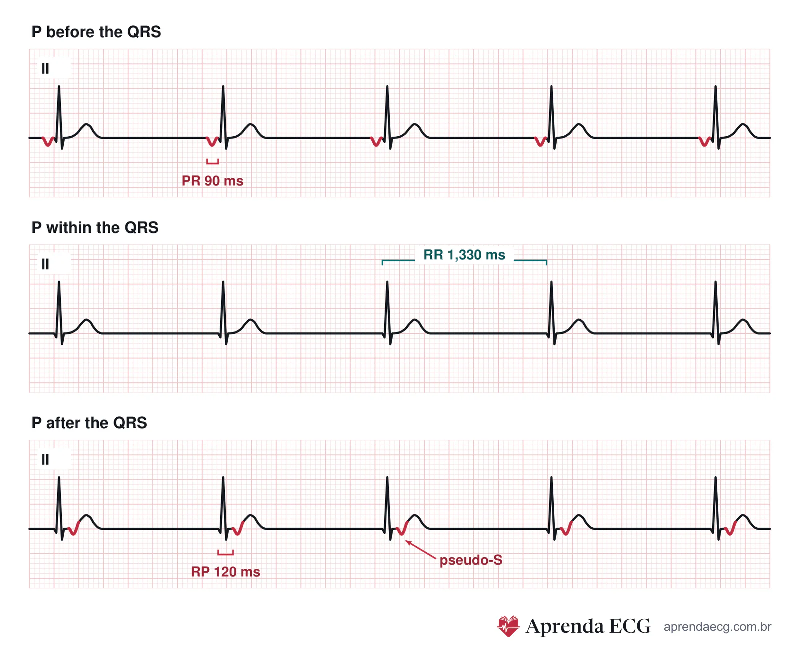
- Before the QRS: PR < 120 ms; with PR ≥ 120 ms, low atrial and junctional rhythms cannot be reliably distinguished.1,2,4,5
- Within the QRS: no visible P wave, smooth baseline between QRS complexes; this is the most common presentation.1,2,4,5
- After the QRS: negative P wave at the end of the QRS complex or in the ST segment: pseudo-S in leads II, III, and aVF and pseudo-r′ in V1 and aVR; the RP interval is variable and may exceed 200 ms.1,2,4,5
- Without retrograde conduction: positive sinus P wave with no fixed relationship to the QRS: AV dissociation, often isorhythmic.1,2,4,5
Naming by Rate
| Rhythm | HR | Usual mechanism | Typical setting |
|---|---|---|---|
| Junctional escape | < 50 bpm | Passive: backup | Sinus bradycardia, sinus arrest, sinoatrial block, third-degree AV block |
| Active (accelerated) junctional rhythm | 50–99 bpm | Active: increased automaticity | Digitalis, ischemia, postoperative state, catecholamines; gradual onset and termination |
| Junctional tachycardia | ≥ 100 bpm | Active | Postoperative state (especially in children), digitalis, ischemia |
- 50 bpm cutoff: many textbooks place the limit between escape and active rhythm at 60 bpm; here at Aprenda ECG the cutoff is 50 bpm, the same as for the normal sinus rate. Between 50 and 60 bpm, the interpretation calls the rhythm active, but the mechanism may be escape.1,3-5
- Active rhythm and dissociation: if retrograde conduction fails, the atria remain in sinus rhythm and AV dissociation develops, with a ventricular rate higher than the atrial rate, often isorhythmic.1,7
Causes
- Physiologic: increased vagal tone, especially during sleep; young people and athletes; up to one third of normal children.1-5,8,9
- Failure of the higher pacemaker or of conduction: sinus node dysfunction, including drug-induced; atrioventricular block.1-5,8,9
- Drugs and catecholamines: beta-blockers, calcium channel blockers, and antiarrhythmic drugs; digitalis (escape with block and active rhythm); isoproterenol and other sympathomimetics.1-5,8,9
- Ischemia and postoperative state: AV nodal ischemia, especially in acute inferior infarction; cardiac surgery (junctional tachycardia especially after valve replacement) and children in the first days after repair of congenital heart disease.1-5,8,9
- Others: myocarditis, acute rheumatic fever, Lyme disease, diphtheria, hyperkalemia, and hypothyroidism.1-5,8,9
Key Points
- Escape is protective: at its usual rate, junctional escape is not a disease in itself; what needs investigation is the failure of the higher pacemaker or of conduction.2,6
- Stability: junctional escape speeds up with exercise, atropine, and catecholamines and is more stable than ventricular escape.3,4
- Active rhythm has a cause: today, infarction and intracardiac surgery are the most common causes; in acute infarction, it appears in up to 10% of patients, more often in inferior than in anterior infarction.1
Pitfalls
- Localizing the focus by the position of the P wave. The position depends on the relative speed of conduction to the atria and to the ventricles, not on how high the focus is in the junction.1,5
- Calling a wide-QRS junctional rhythm ventricular. If bundle branch block was present in sinus rhythm, the junctional QRS is wide; the previous ECG settles it, and a typical bundle branch block morphology, with a rapid initial deflection, favors a junctional origin.1,4
- Mistaking active rhythm for AV nodal reentry. Active junctional rhythm begins and ends gradually, unlike the abrupt onset of atrioventricular nodal reentrant tachycardia (AVNRT).1
- Separating low atrial from junctional rhythm with PR ≥ 120 ms. With a negative P wave in the inferior leads and PR ≥ 120 ms, the two cannot be reliably distinguished.1,2,4,5
Clinical Application
- Interpretation: name the rhythm by rate and describe the position of the P wave and the presence of AV dissociation.5
- Escape: look for what depressed the sinus node or conduction and for reversible causes: drugs, hyperkalemia, hypothyroidism.2
- No intervention: asymptomatic periods of junctional rhythm during sleep, in young people, and in athletes.3,8
Test Yourself
On the 2nd day after valve replacement, the ECG shows a narrow QRS, identical to baseline, regular at 85 bpm. Positive P waves in lead II, at 72 bpm, move in and out of the QRS with no fixed relationship to it. What is the most likely mechanism?
Choose an answer to see the explanation.
Related Topics
References
- Surawicz B, Knilans TK. Chou's Electrocardiography in Clinical Practice. 6ª ed. Philadelphia: Elsevier; 2008.
- Goldberger AL, Goldberger ZD, Shvilkin A. Goldberger's Clinical Electrocardiography: A Simplified Approach. 9a ed. Philadelphia: Elsevier; 2018.
- Scheffer MK, De Marchi MFN, Alencar Neto JN, Felicioni SP, editores. Eletrocardiograma de A a Z. 1a ed. Barueri (SP): Manole; 2024.
- Das MK, Zipes DP. Electrocardiography of Arrhythmias: A Comprehensive Review (A Companion to Cardiac Electrophysiology: From Cell to Bedside). 2a ed. Philadelphia: Elsevier; 2022.
- Samesima N, God EG, Kruse JCL, Leal MG, Pinho C, França FFAC, et al. Brazilian Society of Cardiology Guidelines on the Analysis and Issuance of Electrocardiographic Reports - 2022. Arq Bras Cardiol. 2022;119(4):638-680.
- Bayés de Luna A, Fiol-Sala M, Bayés-Genís A, Baranchuk A. Clinical Electrocardiography: A Textbook. 5a ed. Hoboken (NJ): Wiley; 2022.
- Issa ZF, Miller JM, Zipes DP. Clinical Arrhythmology and Electrophysiology: A Companion to Braunwald's Heart Disease. 3ª ed. Philadelphia: Elsevier; 2019.
- Kusumoto FM, Schoenfeld MH, Barrett C, Edgerton JR, Ellenbogen KA, Gold MR, et al. 2018 ACC/AHA/HRS Guideline on the Evaluation and Management of Patients With Bradycardia and Cardiac Conduction Delay: A Report of the American College of Cardiology/American Heart Association Task Force on Clinical Practice Guidelines and the Heart Rhythm Society. J Am Coll Cardiol. 2019;74(7):e51-e156.
- Sharma S, Drezner JA, Baggish A, Papadakis M, Wilson MG, Prutkin JM, et al. International Recommendations for Electrocardiographic Interpretation in Athletes. J Am Coll Cardiol. 2017;69(8):1057-1075.