Ectopic atrial rhythm arises from an atrial focus outside the sinus node. It is a common finding on the resting ECG and Holter monitoring, especially during sleep, in athletes, and in children, and most of the time it has no consequence.1-4
Criteria
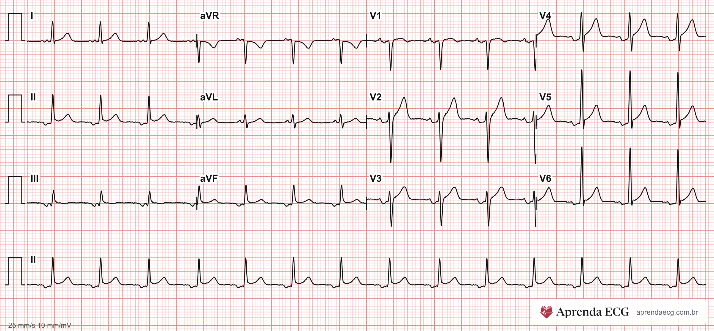
- P wave: morphology different from sinus and, usually, an axis outside the sinus range (0° to +90°, with a positive P wave in leads I, II, and aVF); a focus close to the sinus node produces a P wave almost identical to the sinus P wave.1,5-7
- P–QRS relationship: one P wave before each QRS complex.1,8
- PR interval: usually ≥ 120 ms.1,4,5,8,9
- Heart rate: 50 to 99 bpm.1,4,5,8,9
| Rhythm | HR | P wave | PR interval |
|---|---|---|---|
| Ectopic atrial rhythm | 50–99 bpm | One morphology, different from sinus; one P wave per QRS complex | Usually ≥ 120 ms |
| Atrial escape rhythm (ectopic atrial bradycardia) | < 50 bpm | Same as above | Usually ≥ 120 ms |
| Wandering atrial pacemaker | < 100 bpm | ≥ 3 different morphologies; one P wave per QRS complex | ≥ 3 different values |
| Atrial tachycardia (focal or multifocal) | ≥ 100 bpm | One or several morphologies | Variable |
Mechanism
- Passive form (escape): the sinus node slows, because of vagal tone or disease, and a latent atrial pacemaker takes control.1,8,10
- Active form: the atrial focus fires faster than the sinus node and overrides it.1,8,10
- Course: usually transient, and easiest to recognize when the same tracing shows the sinus P wave for comparison.1,8
Wandering Atrial Pacemaker
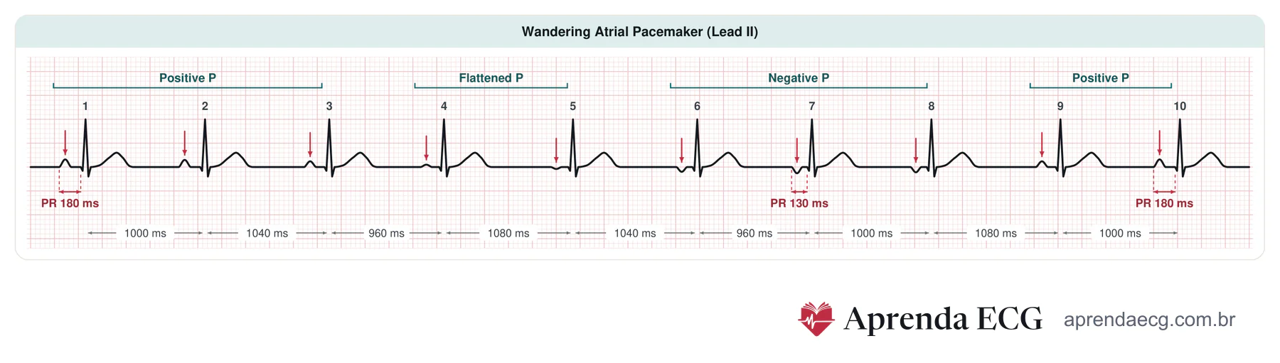
- Criteria: at least 3 P-wave morphologies and 3 different PR intervals, changing gradually over several beats; variable PP and RR intervals, usually one P wave per QRS complex, HR below 100 bpm.5,8,9
- With HR ≥ 100 bpm: the same pattern is multifocal atrial tachycardia.5,9,10
- Significance: usually a physiologic variant related to vagal tone; it also occurs in sinus node dysfunction, drug toxicity, and structural heart disease.7,9,10
Differential Diagnosis
- Sinus rhythm and sinus arrhythmia: positive P wave in leads I, II, and aVF; in sinus arrhythmia, the P wave changes little and follows respiration.1,5,7,10,11
- Junctional rhythm: negative P wave in the inferior leads with PR < 120 ms, or P wave within or after the QRS complex.1,5,7,10,11
- Focal atrial tachycardia: same nonsinus P wave, with HR ≥ 100 bpm.1,5,7,10,11
- Premature atrial complexes: premature, isolated P waves over an underlying sinus rhythm.1,5,7,10,11
- Arm electrode reversal and dextrocardia: negative P wave and QRS complex in lead I; with reversal, normal precordial leads; with dextrocardia, poor R-wave progression.1,5,7,10,11
- Right arm–left leg electrode reversal: negative P wave, QRS complex, and T wave in leads I, II, III, and aVF and positive in aVR; in an atrial focus, only the P wave changes.1,5,7,10,11
Key Points
- Rate ranges: with HR < 50 bpm, it is atrial escape (ectopic atrial bradycardia); with HR ≥ 100 bpm, atrial tachycardia.1,4,5,8,9
- Benign in most cases: with a normal HR and no symptoms, it is almost always benign; it is a normal finding during sleep, in athletes, and in children.2,3,9
- Athletes: junctional escape rhythm or wandering pacemaker appears in up to 8% at rest, and sinus rhythm returns with exertion.2,3,9,10
Pitfalls
- Calling a low atrial focus junctional rhythm. Both produce a negative P wave in the inferior leads; the PR interval separates them: usually ≥ 120 ms in the atrial focus, < 120 ms or P wave within or after the QRS complex in junctional rhythm.1,5,6,8
- Calling electrode reversal an ectopic rhythm. If the P wave, QRS complex, and T wave change together in the limb leads, think of reversal; in an atrial focus, only the P wave changes.1,5,7,10,11
- Missing sinus node dysfunction. Below 50 bpm while awake, ectopic atrial rhythm may be the manifestation of a subsidiary pacemaker taking control.4
- Mistaking wandering pacemaker for atrial fibrillation. The rhythm is irregular, but the P waves are distinct and separated by an isoelectric baseline.8,9
Test Yourself
An ECG shows distinct P waves of four different morphologies, separated by an isoelectric baseline, one before each QRS complex, with three different PR intervals, an irregular RR, a narrow QRS complex, and a heart rate of 112 bpm. What is the rhythm diagnosis?
Choose an answer to see the explanation.
Related Topics
References
- Surawicz B, Knilans TK. Chou's Electrocardiography in Clinical Practice. 6ª ed. Philadelphia: Elsevier; 2008.
- Drezner JA, Sharma S, Baggish A, Papadakis M, Wilson MG, Prutkin JM, et al. International criteria for electrocardiographic interpretation in athletes: Consensus statement. Br J Sports Med. 2017;51(9):704-731.
- Drago F, Battipaglia I, Di Mambro C. Neonatal and Pediatric Arrhythmias: Clinical and Electrocardiographic Aspects. Card Electrophysiol Clin. 2018;10(2):397-412.
- Kusumoto FM, Schoenfeld MH, Barrett C, Edgerton JR, Ellenbogen KA, Gold MR, et al. 2018 ACC/AHA/HRS Guideline on the Evaluation and Management of Patients With Bradycardia and Cardiac Conduction Delay: A Report of the American College of Cardiology/American Heart Association Task Force on Clinical Practice Guidelines and the Heart Rhythm Society. J Am Coll Cardiol. 2019;74(7):e51-e156.
- Samesima N, God EG, Kruse JCL, Leal MG, Pinho C, França FFAC, et al. Brazilian Society of Cardiology Guidelines on the Analysis and Issuance of Electrocardiographic Reports - 2022. Arq Bras Cardiol. 2022;119(4):638-680.
- Pastore CA, Samesima N, Tobias NMMO, Pereira Filho HG. Eletrocardiografia Atual: Curso do Serviço de Eletrocardiografia do InCor. 3a ed. São Paulo: Atheneu; 2016.
- Das MK, Zipes DP. Electrocardiography of Arrhythmias: A Comprehensive Review (A Companion to Cardiac Electrophysiology: From Cell to Bedside). 2a ed. Philadelphia: Elsevier; 2022.
- Scheffer MK, De Marchi MFN, Alencar Neto JN, Felicioni SP, editores. Eletrocardiograma de A a Z. 1a ed. Barueri (SP): Manole; 2024.
- Issa ZF, Miller JM, Zipes DP. Clinical Arrhythmology and Electrophysiology: A Companion to Braunwald's Heart Disease. 3ª ed. Philadelphia: Elsevier; 2019.
- Goldberger AL, Goldberger ZD, Shvilkin A. Goldberger's Clinical Electrocardiography: A Simplified Approach. 9a ed. Philadelphia: Elsevier; 2018.
- Paul A, Jacob JR. Electrocardiographic lead reversals. Indian Pacing Electrophysiol J. 2023;23(6):205-213.