

AF is defined by chaotic, disorganized atrial electrical activity with absent P waves, producing fine, coarse or mixed irregularities in the baseline (f waves). The atrial rate ranges from 450 to 700 cycles per minute, with a variable ventricular response.
Electrocardiographic Criteria
- Absence of P waves
- Irregular RR intervals
- The baseline may be isoelectric or show fine or coarse irregularities (f waves)
Classification
The current classification of AF distinguishes five patterns based on the presentation, duration and spontaneous termination of arrhythmia episodes.
- First-diagnosed AF: AF not diagnosed before, regardless of its duration or the presence/severity of AF-related symptoms
- Paroxysmal AF: AF that terminates spontaneously or with intervention within 7 days of onset
- Persistent AF: AF continuously sustained for more than 7 days, including episodes terminated by cardioversion (drugs or electrical cardioversion) after ≥ 7 days
- Long-standing persistent AF: continuous AF lasting > 12 months when it is decided to adopt a rhythm control strategy
- Permanent AF: AF accepted by the patient and the physician, with no further attempts to restore/maintain sinus rhythm. The term should not be used in the context of a rhythm control strategy (antiarrhythmic therapy or ablation). If a rhythm control strategy is adopted, the arrhythmia should be reclassified as “long-standing persistent AF”
Ashman Phenomenon
The Ashman phenomenon is a type of rate-dependent intraventricular conduction disturbance. It occurs in a beat that follows a short RR interval after a long RR interval (long-short cycle sequences), due to the increased refractory period of the conduction system. An aberrant beat with RBBB morphology is usually observed.
ECG in Atrial Fibrillation
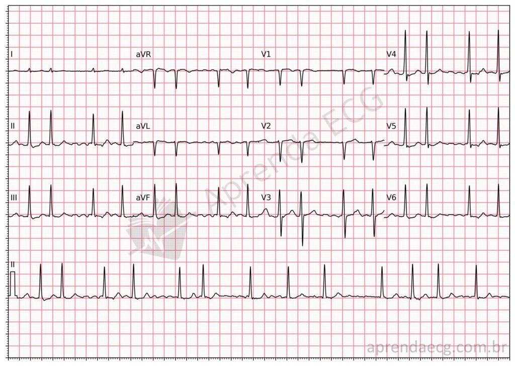
Atrial fibrillation. Irregular rhythm with absent P waves. The baseline irregularities are caused by f waves.
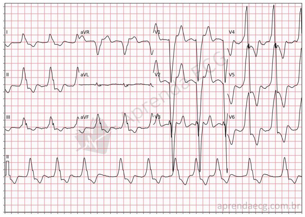
Atrial fibrillation. Example of AF with LBBB; note the irregularity between the QRS complexes and the absence of P waves on the ECG.
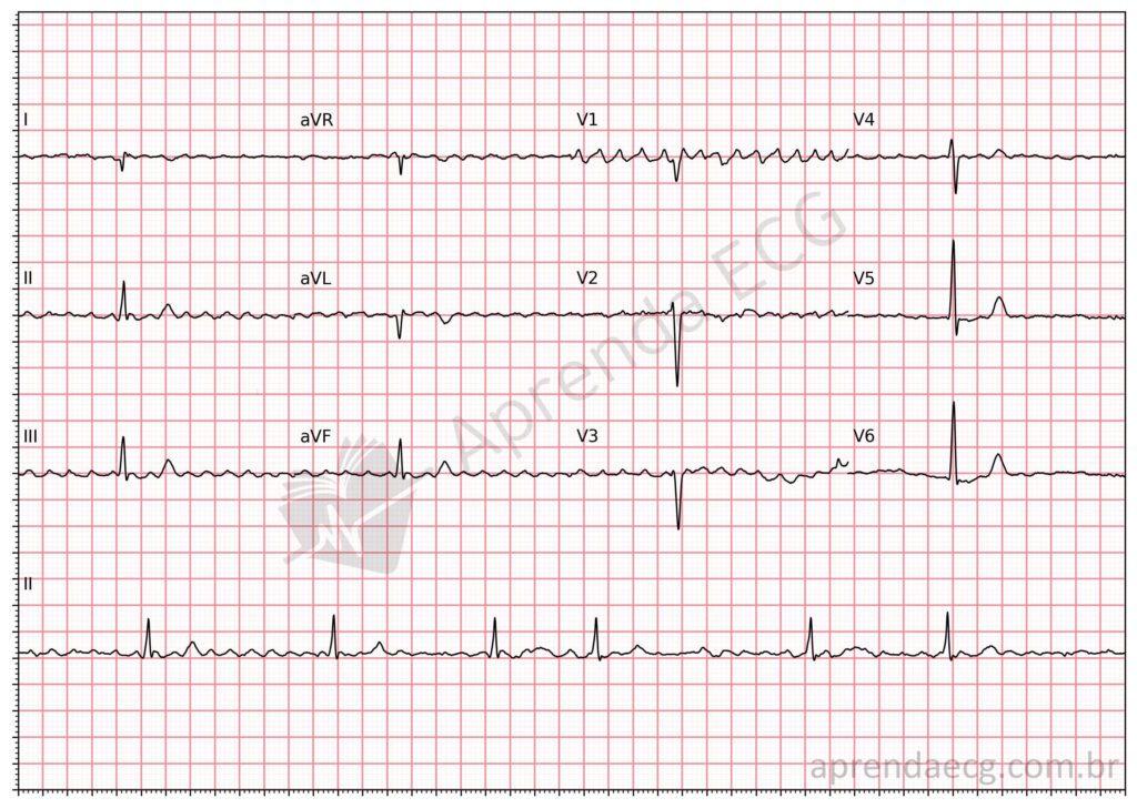
Atrial fibrillation. Example of AF with a slow ventricular response. The f waves are visible on the tracing, producing coarse oscillations.
Further Reading
Related Topics
References
- Samesima N, God EG, Kruse JCL, Leal MG, et al. Diretriz da Sociedade Brasileira de Cardiologia sobre a Análise e Emissão de Laudos Eletrocardiográficos – 2022. Arq Bras Cardiol. 2022;93(3 Suppl 2):2–19.
- Hindricks G, Potpara T, Dagres N, Bax JJ, et al. 2020 ESC Guidelines for the diagnosis and management of atrial fibrillation developed in collaboration with the European Association for Cardio-Thoracic Surgery (EACTS). Eur Heart J. 2021;42(5):373–498.
- Campelo RT, Armaganijan L, Moreira DAR, Scheffer MK, et al. Amplitude das Ondas F como Preditor de Tromboembolismo e de Sucesso da Cardioversão Elétrica em Pacientes com Fibrilação Atrial Persistente. Arq Bras Cardiol. 2022;
- Gouaux JL, Ashman R. Auricular fibrillation with aberration simulating ventricular paroxysmal tachycardia. Am Heart J. 1947;34(3):366–73.


