Accelerated idioventricular rhythm (AIVR) is an ectopic ventricular rhythm faster than idioventricular escape and slower than ventricular tachycardia (VT); it is the most frequent arrhythmia during reperfusion of acute myocardial infarction (AMI).1-3 Recognizing it avoids errors in both directions: treating a benign, self-limited rhythm as VT or as complete atrioventricular block, or calling a slow VT that needs different management AIVR.1
Criteria
- QRS: wide (≥ 120 ms), not preceded by a conducted P wave.4-6
- Sequence: three or more consecutive complexes, usually monomorphic and with regular RR.4-6
- Heart rate (HR): 50 to 110 bpm, the range adopted here at Aprenda ECG; other sources give ranges between 50–100 and 50–130 bpm (usually 70 to 85 bpm).4-9
- Onset and termination: gradual.4-6
- Fusion and capture beats: frequent.4-6
Mechanism
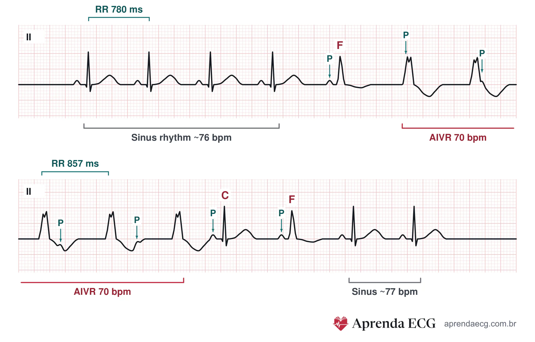
- Competition, not backup: unlike escape, AIVR competes with the underlying rhythm. It appears when the sinus rate falls below the rate of the focus, which is why it usually emerges with sinus bradycardia, and it disappears when the sinus rate speeds up again.3-7
- Abnormal automaticity: acceleration of diastolic depolarization (phase 4) in His-Purkinje fibers or ventricular myocardium; in digitalis toxicity and in some situations of acute ischemia, triggered activity has been proposed.1,4,5,10,11
- Unprotected focus: unlike parasystole, it is discharged by the sinus rhythm whenever the sinus rate becomes faster.3,7
- Fusion, capture, and dissociation: with similar rates, the two pacemakers compete for the ventricles; fusion and capture beats are much more frequent than in VT and cluster at the beginning and end of episodes, and isorhythmic dissociation is common.1,3,5,9
Reperfusion in Infarction
- Incidence: 8% to 36% of AMIs in the early series, predominantly in the first 12 to 24 hours; during primary angioplasty in ST-elevation myocardial infarction (STEMI), it was the most common arrhythmia (42% of 503 patients).2,3,10
- After streptokinase: AIVR in the 2 hours after the infusion had a sensitivity of 20% and a specificity of 98% for successful thrombolysis (positive likelihood ratio of 8.9 and negative of 0.82, calculated from the study); its absence barely changes the probability.12
- In primary angioplasty: a nonspecific marker; it did not distinguish TIMI 2 from TIMI 3 flow and was associated with larger infarction, with no difference in mortality. It suggests that the artery has opened, but not that reperfusion was complete.1,2,11
Differential Diagnosis
| Feature | Idioventricular escape | AIVR | Monomorphic VT |
|---|---|---|---|
| HR | < 40 bpm | 50–110 bpm | > 100 bpm, usually > 120 bpm |
| Onset | Late, after a pause or failure of the higher rhythm | Gradual, when the sinus rate falls below that of the focus | Abrupt, usually triggered by an early premature beat |
| Relationship with the underlying rhythm | Replaces an inhibited or blocked rhythm (complete atrioventricular block, sinus arrest) | Competes with it | Independent |
| Fusion and capture beats | Possible | Frequent | Rare |
| Course | Persists while the higher rhythm fails | Self-limited | May degenerate and cause instability |
Causes
- Coronary artery disease: reperfusion in AMI (thrombolysis, angioplasty, or spontaneous), the most common cause; acute ischemia.1,3-5,13
- Drugs and toxins: digitalis toxicity, cocaine, inhaled anesthetics (halothane, desflurane).1,3-5,13
- Others: hypokalemia and hyperkalemia; structural heart disease and myocarditis; post-resuscitation.1,3-5,13
- Normal heart: adolescents and athletes (vagal predominance); 0.3% of healthy volunteers on ambulatory monitoring.1,3-5,13
Key Points
- Benign in most cases: usually well tolerated and self-limited; the prognosis is that of the underlying disease.1,3,11
- Rarely degenerates: into VT or ventricular fibrillation, although paroxysmal VT may coexist in the same patient.1,3
Pitfalls
- Deciding by HR alone. The AIVR range overlaps with that of some slow VTs; the mode of onset, the relationship with the underlying rhythm, and the clinical context also count.1
- Ignoring the beat that starts the rhythm. AIVR initiated by an early premature beat, rather than by a late escape beat, is more often associated with fast ventricular tachyarrhythmias.7
- Calling a wide-QRS junctional rhythm ventricular. Junctional rhythm with bundle branch block or aberrancy can mimic a slow ventricular rhythm, sometimes with no possible distinction on the ECG; fusion and capture beats support a ventricular origin.1,3
- Using AIVR as proof of complete reperfusion. In primary angioplasty, it did not distinguish TIMI 2 from TIMI 3 flow.2,11
Clinical Application
- During reperfusion: monitoring and correcting the cause are enough.4,10
- Severe ventricular dysfunction: loss of atrioventricular synchrony can cause hypotension.1
- Hemodynamic instability: speeding up the sinus rhythm with atropine or atrial pacing restores synchrony; antiarrhythmic drugs are rarely needed and can cause hemodynamic deterioration, because they suppress the rhythm that is maintaining the HR.1,13
Test Yourself
A patient with severe ventricular dysfunction develops accelerated idioventricular rhythm at 68 bpm, with hypotension attributed to loss of atrioventricular synchrony. Which measure addresses the mechanism of the instability?
Choose an answer to see the explanation.
Related Topics
References
- Ricardo Pérez Riera A, Barbosa Barros R, Henrique Kawatake de Souza D, Daminello Raimundo R, Sclarovsky S, Nikus K. Accelerated idioventricular rhythm in the adult: an update. Journal of Human Growth and Development. 2025;35(1):158-165.
- Terkelsen CJ, Sørensen JT, Kaltoft AK, Nielsen SS, Thuesen L, Bøtker HE, et al. Prevalence and significance of accelerated idioventricular rhythm in patients with ST-elevation myocardial infarction treated with primary percutaneous coronary intervention. Am J Cardiol. 2009;104(12):1641-6.
- Surawicz B, Knilans TK. Chou's Electrocardiography in Clinical Practice. 6ª ed. Philadelphia: Elsevier; 2008.
- Scheffer MK, De Marchi MFN, Alencar Neto JN, Felicioni SP, editores. Eletrocardiograma de A a Z. 1a ed. Barueri (SP): Manole; 2024.
- Riera ARP, Barros RB, de Sousa FD, Baranchuk A. Accelerated Idioventricular Rhythm: History and Chronology of the Main Discoveries. Indian Pacing Electrophysiol J. 2010;10(1):40-8.
- Samesima N, God EG, Kruse JCL, Leal MG, Pinho C, França FFAC, et al. Brazilian Society of Cardiology Guidelines on the Analysis and Issuance of Electrocardiographic Reports - 2022. Arq Bras Cardiol. 2022;119(4):638-680.
- Goldberger AL, Goldberger ZD, Shvilkin A. Goldberger's Clinical Electrocardiography: A Simplified Approach. 9a ed. Philadelphia: Elsevier; 2018.
- Pastore CA, Samesima N, Tobias NMMO, Pereira Filho HG. Eletrocardiografia Atual: Curso do Serviço de Eletrocardiografia do InCor. 3a ed. São Paulo: Atheneu; 2016.
- Bayés de Luna A, Fiol-Sala M, Bayés-Genís A, Baranchuk A. Clinical Electrocardiography: A Textbook. 5a ed. Hoboken (NJ): Wiley; 2022.
- Issa ZF, Miller JM, Zipes DP. Clinical Arrhythmology and Electrophysiology: A Companion to Braunwald's Heart Disease. 3ª ed. Philadelphia: Elsevier; 2019.
- Bonnemeier H, Ortak J, Wiegand UKH, Eberhardt F, Bode F, Schunkert H, et al. Accelerated idioventricular rhythm in the post-thrombolytic era: incidence, prognostic implications, and modulating mechanisms after direct percutaneous coronary intervention. Ann Noninvasive Electrocardiol. 2005;10(2):179-87.
- Khan A, Nadeem S, Kokane H, Thummar A, Lokhandwala Y, Mahajan AU, et al. Is accelerated idioventricular rhythm a good marker for reperfusion after streptokinase? Indian Heart J. 2016;68(3):302-5.
- Gildea TH, Levis JT. ECG Diagnosis: Accelerated Idioventricular Rhythm. Perm J. 2018;22(2):17-173.