

Today we are going to learn how to identify the rhythm on the ECG.
Identifying the rhythm means determining whether it is sinus in origin, whether it is a wide or narrow QRS tachycardia, whether it originates from some other focus within the atria (ectopic atrial rhythm), or whether it is an escape rhythm.
The most important point here is to identify the presence of atrial electrical activity and determine whether or not it originated in the sinus node.
So let's start by first learning how to identify a sinus rhythm.
How to Identify Sinus Rhythm
After Step 0 of ECG analysis, which consists of identifying the characteristics of our patient and the technical quality of the tracing, Step 1 is the analysis of the cardiac rhythm.
The normal rhythm, or sinus rhythm, is the one that originates in the heart's dominant pacemaker, the sinus node. This structure is located in the upper portion of the right atrium. Therefore, electrical impulses that originate in this region spread to the remaining right and left atrial cells, so that the activation vector points downward and to the left.
Note that the result of this is the recording of the P wave on the ECG.
Since the vector points downward and to the left, its resultant will point to approximately +60°, which is exactly where lead II is located. For this reason, the P wave is usually most easily seen and has the greatest amplitude in this lead.
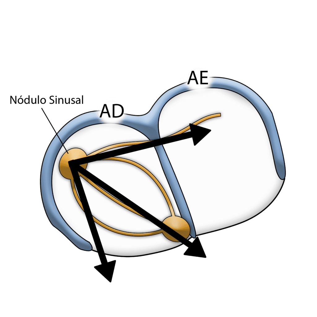
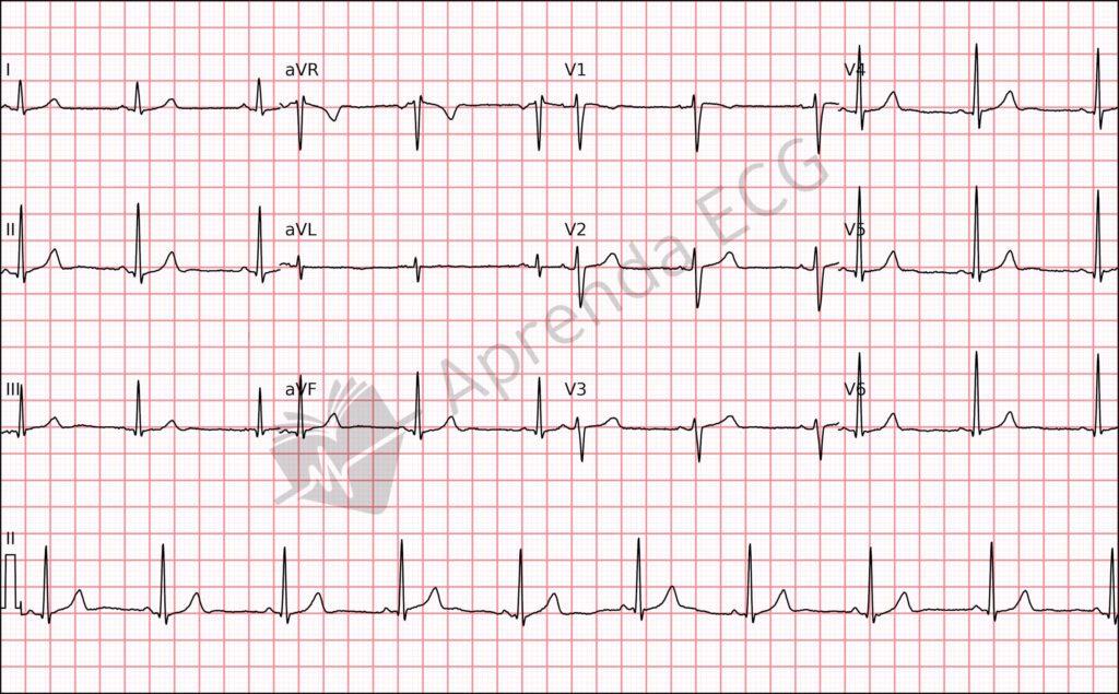
Based on this knowledge, to say that a rhythm is sinus we need to find the following criteria:
- A positive P wave in I, II, and aVF
- A biphasic (plus-minus) or negative P wave in V1
- And a positive P wave in V6
Ectopic Atrial and Junctional Rhythms
An ectopic atrial rhythm can originate in any region of the atria and will therefore produce P waves with morphologies different from the sinus P wave we saw earlier. They do not meet the criteria for sinus rhythm.
Entirely positive P waves in V1 are not expected and suggest an ectopic atrial rhythm, usually from a focus in the left atrium.
Another situation is junctional rhythm, in which the electrical impulse originates in the AV node, and we may find negative P waves in the inferior leads (II, III, and aVF) with a short PR interval; or absent P waves; or a negative P wave in the inferior leads after the QRS complex.
Escape Rhythms
Escape rhythms are safety mechanisms of the heart's electrical conduction system, activated when the sinus node fails to generate impulses or when their conduction is blocked.
These rhythms arise from secondary automatic foci, such as the atrioventricular node (junctional escape) or the ventricular myocardium (ventricular escape).
Let's look at their criteria:
- Junctional Escape Rhythm
- Backup or replacement rhythm originating in the AV node, with QRS complexes identical to or slightly different from those of sinus origin
- HR usually < 50 bpm
- Ventricular Escape Rhythm
- Rhythm originating in the ventricles that takes over when anatomically higher rhythms have been suppressed or blocked
- Wide QRS (> 120 ms) and HR usually < 40 bpm
Irregular Rhythms
Among irregular rhythms there are a few possibilities: first, rule out whether the irregularity is caused by premature beats or pauses. Then assess whether or not atrial activity is present.
The absence of P waves with an isoelectric baseline or even small baseline oscillations corresponds to atrial fibrillation.
The presence of P waves with variable morphologies and PR intervals strongly suggests multifocal atrial tachycardia.
On the other hand, the presence of atrial flutter F waves or P waves with morphologies different from the sinus P wave points to flutter with variable AV conduction or atrial tachycardia with variable AV conduction, respectively.
Narrow QRS Tachycardias
A narrow QRS tachycardia is defined as a rhythm with a rate > 100 bpm and a QRS < 120 ms.
In this situation, the rhythm is probably supraventricular in origin.
It may be sinus tachycardia, focal atrial tachycardia, AV nodal reentrant tachycardia, atrial flutter… among many other supraventricular tachycardias, in which the ECG will not always point to a definitive mechanism.
The differential diagnosis of supraventricular tachycardias on the ECG is a complex topic, and I recommend that you explore our ECG Library to learn the criteria for each of them.
Wide QRS Tachycardias
If the QRS is prolonged, > 120 ms in duration, the rhythm may be supraventricular with aberrant conduction through one of the bundle branches or fascicles of the His-Purkinje system, or it may be ventricular tachycardia.
What helps us in this scenario is applying the wide QRS tachycardia algorithms, of which the Brugada Algorithm is the best known and most widely used.
There are other, less common causes of wide QRS tachycardia. In the tools section you will find them along with the main algorithms used in practice.
Practice Makes Perfect
As we have seen, rhythm analysis ranges from identifying sinus rhythm to the differential diagnosis between supraventricular and ventricular tachycardias. Learning ECG takes time, but with practice you will be able to identify the different rhythms you encounter and become a reference in ECG analysis.
Our ECG Library is designed to help you in this process, providing examples of real tracings and easy access to electrocardiographic criteria.


